 Movie
Movie Controller


+ Open data

- Basic information
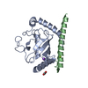
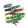
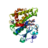
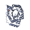
| Entry | Database: PDB / ID: 4jqu | ||||||
|---|---|---|---|---|---|---|---|
| Title | Crystal structure of Ubc7p in complex with the U7BR of Cue1p | ||||||
 Components Components |
| ||||||
 Keywords Keywords | LIGASE/PROTEIN BINDING / Ubc7p:U7BR complex / ligase-activator complex / LIGASE-PROTEIN BINDING complex | ||||||
| Function / homology |  Function and homology information Function and homology informationCUE1-UBC7 ubiquitin-conjugating enzyme complex / establishment of protein localization to endoplasmic reticulum membrane / Doa10p ubiquitin ligase complex / Synthesis of active ubiquitin: roles of E1 and E2 enzymes / Hrd1p ubiquitin ligase ERAD-L complex / fungal-type cell wall organization / ubiquitin-protein transferase activator activity / Antigen processing: Ubiquitination & Proteasome degradation /  E2 ubiquitin-conjugating enzyme / : ...CUE1-UBC7 ubiquitin-conjugating enzyme complex / establishment of protein localization to endoplasmic reticulum membrane / Doa10p ubiquitin ligase complex / Synthesis of active ubiquitin: roles of E1 and E2 enzymes / Hrd1p ubiquitin ligase ERAD-L complex / fungal-type cell wall organization / ubiquitin-protein transferase activator activity / Antigen processing: Ubiquitination & Proteasome degradation / E2 ubiquitin-conjugating enzyme / : / ERAD pathway / ubiquitin conjugating enzyme activity / response to cadmium ion / ubiquitin binding / ubiquitin-protein transferase activity / chromatin organization / ubiquitin-dependent protein catabolic process / protein ubiquitination / endoplasmic reticulum membrane / endoplasmic reticulum / mitochondrion / ATP binding / cytosol E2 ubiquitin-conjugating enzyme / : ...CUE1-UBC7 ubiquitin-conjugating enzyme complex / establishment of protein localization to endoplasmic reticulum membrane / Doa10p ubiquitin ligase complex / Synthesis of active ubiquitin: roles of E1 and E2 enzymes / Hrd1p ubiquitin ligase ERAD-L complex / fungal-type cell wall organization / ubiquitin-protein transferase activator activity / Antigen processing: Ubiquitination & Proteasome degradation / E2 ubiquitin-conjugating enzyme / : / ERAD pathway / ubiquitin conjugating enzyme activity / response to cadmium ion / ubiquitin binding / ubiquitin-protein transferase activity / chromatin organization / ubiquitin-dependent protein catabolic process / protein ubiquitination / endoplasmic reticulum membrane / endoplasmic reticulum / mitochondrion / ATP binding / cytosolSimilarity search - Function | ||||||
| Biological species |  Saccharomyces cerevisiae (brewer's yeast) Saccharomyces cerevisiae (brewer's yeast) | ||||||
| Method | X-RAY DIFFRACTION / SYNCHROTRON / MOLECULAR REPLACEMENT / molecular replacement / Resolution: 1.81 Å | ||||||
 Authors Authors | Liang, Y.-H. / Metzger, M.B. / Weissman, A.M. / Ji, X. | ||||||
 Citation Citation | Journal: Mol.Cell / Year: 2013 Title: A Structurally Unique E2-Binding Domain Activates Ubiquitination by the ERAD E2, Ubc7p, through Multiple Mechanisms. Authors: Metzger, M.B. / Liang, Y.H. / Das, R. / Mariano, J. / Li, S. / Li, J. / Kostova, Z. / Byrd, R.A. / Ji, X. / Weissman, A.M. | ||||||
| History |
|
- Structure visualization
| Structure viewer | Molecule: MolmilJmol/JSmol |
|---|
- Downloads & links
-Download
| PDBx/mmCIF format | 4jqu.cif.gz | 63.8 KB | Display | PDBx/mmCIF format |
|---|---|---|---|---|
| PDB format | pdb4jqu.ent.gz | 45.3 KB | Display | PDB format |
| PDBx/mmJSON format | 4jqu.json.gz | Tree view | PDBx/mmJSON format | |
| Others |  Other downloads Other downloads |
-Validation report
| Arichive directory | https://data.pdbj.org/pub/pdb/validation_reports/jq/4jquftp://data.pdbj.org/pub/pdb/validation_reports/jq/4jqu | HTTPS FTP |
|---|
-Related structure data
| Related structure data |  2uczS S: Starting model for refinement |
|---|---|
| Similar structure data |


-Links
 PDBj
PDBj


- Assembly
| Deposited unit | 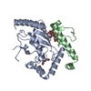
| ||||||||
|---|---|---|---|---|---|---|---|---|---|
| 1 |
| ||||||||
| Unit cell |
|
-Components
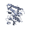
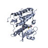
| #1: Protein | Mass: 18764.441 Da / Num. of mol.: 1 Source method: isolated from a genetically manipulated source Source: (gene. exp.) Saccharomyces cerevisiae (brewer's yeast)Strain: ATCC 204508 / S288c / Gene: QRI8, UBC7, Ubc7p, YM9711.12, YMR022W / Plasmid: pGEX / Production host:  Escherichia coli (E. coli) / Strain (production host): BL21(star) / References: UniProt: Q02159, ubiquitin-protein ligase Escherichia coli (E. coli) / Strain (production host): BL21(star) / References: UniProt: Q02159, ubiquitin-protein ligase | ||
|---|---|---|---|
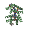
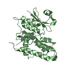
| #2: Protein | Mass: 6485.444 Da / Num. of mol.: 1 / Fragment: U7BR (UNP residues 151-203) Source method: isolated from a genetically manipulated source Source: (gene. exp.) Saccharomyces cerevisiae (brewer's yeast)Strain: ATCC 204508 / S288c / Gene: CUE1, Cue1p, KIS4, YM8156.06, YMR264W / Plasmid: pGEX / Production host: Escherichia coli (E. coli) / Strain (production host): BL21(star) / References: UniProt: P38428 | ||
| #3: Chemical | ChemComp-BTB / Bis-tris methane  Mass: 209.240 Da / Num. of mol.: 1 / Source method: obtained synthetically / Formula: C8H19NO5 / Comment: pH buffer*YM Mass: 209.240 Da / Num. of mol.: 1 / Source method: obtained synthetically / Formula: C8H19NO5 / Comment: pH buffer*YM | ||
| #4: Chemical | Diethylene glycol  Mass: 106.120 Da / Num. of mol.: 2 / Source method: obtained synthetically / Formula: C4H10O3 Mass: 106.120 Da / Num. of mol.: 2 / Source method: obtained synthetically / Formula: C4H10O3#5: Water | ChemComp-HOH / | Water Mass: 18.015 Da / Num. of mol.: 136 / Source method: isolated from a natural source / Formula: H2O Mass: 18.015 Da / Num. of mol.: 136 / Source method: isolated from a natural source / Formula: H2O |
-Experimental details
-Experiment
| Experiment | Method: X-RAY DIFFRACTION / Number of used crystals: 1 |
|---|
- Sample preparation
| Crystal | Density Matthews: 2.15 Å3/Da / Density % sol: 42.8 % |
|---|---|
| Crystal grow | Temperature: 293 K / Method: vapor diffusion, sitting drop / pH: 6.5 Details: 25% PEG3350, 0.2 M ammonium acetate, 0.1 M Bis-Tris, pH 6.5, VAPOR DIFFUSION, SITTING DROP, temperature 293K |
-Data collection
| Diffraction | Mean temperature: 100 K | |||||||||||||||||||||||||||||||||||||||||||||||||||||||||||||||||||||||||||||
|---|---|---|---|---|---|---|---|---|---|---|---|---|---|---|---|---|---|---|---|---|---|---|---|---|---|---|---|---|---|---|---|---|---|---|---|---|---|---|---|---|---|---|---|---|---|---|---|---|---|---|---|---|---|---|---|---|---|---|---|---|---|---|---|---|---|---|---|---|---|---|---|---|---|---|---|---|---|---|
| Diffraction source | Source: SYNCHROTRON / Site: APS  / Beamline: 22-BM / Wavelength: 1 Å / Beamline: 22-BM / Wavelength: 1 Å | |||||||||||||||||||||||||||||||||||||||||||||||||||||||||||||||||||||||||||||
| Detector | Type: MARMOSAIC 225 mm CCD / Detector: CCD / Date: Nov 23, 2009 | |||||||||||||||||||||||||||||||||||||||||||||||||||||||||||||||||||||||||||||
| Radiation | Monochromator: Rosenbaum-Rock double-crystal Si(220) / Protocol: SINGLE WAVELENGTH / Monochromatic (M) / Laue (L): M / Scattering type: x-ray | |||||||||||||||||||||||||||||||||||||||||||||||||||||||||||||||||||||||||||||
| Radiation wavelength | Wavelength: 1 Å / Relative weight: 1 | |||||||||||||||||||||||||||||||||||||||||||||||||||||||||||||||||||||||||||||
| Reflection | Resolution: 1.81→40 Å / Num. all: 20415 / Num. obs: 20415 / % possible obs: 98.7 % / Observed criterion σ(F): -6 / Observed criterion σ(I): -3 / Redundancy: 7.5 % / Rmerge(I) obs: 0.063 / Χ2: 1.008 / Net I/σ(I): 27 | |||||||||||||||||||||||||||||||||||||||||||||||||||||||||||||||||||||||||||||
| Reflection shell | Diffraction-ID: 1
|
-Phasing
| Phasing | Method: molecular replacement |
|---|
- Processing
| Software |
| ||||||||||||||||||||||||||||||||||||||||||||||||||||||||
|---|---|---|---|---|---|---|---|---|---|---|---|---|---|---|---|---|---|---|---|---|---|---|---|---|---|---|---|---|---|---|---|---|---|---|---|---|---|---|---|---|---|---|---|---|---|---|---|---|---|---|---|---|---|---|---|---|---|
| Refinement | Method to determine structure: MOLECULAR REPLACEMENT Starting model: PDB ENTRY 2UCZ Resolution: 1.81→33.467 Å / Occupancy max: 1 / Occupancy min: 0.24 / FOM work R set: 0.854 / SU ML: 0.22 / σ(F): 1.34 / Phase error: 21.13 / Stereochemistry target values: ML
| ||||||||||||||||||||||||||||||||||||||||||||||||||||||||
| Solvent computation | Shrinkage radii: 0.9 Å / VDW probe radii: 1.11 Å / Solvent model: FLAT BULK SOLVENT MODEL / Bsol: 62.188 Å2 / ksol: 0.365 e/Å3 | ||||||||||||||||||||||||||||||||||||||||||||||||||||||||
| Displacement parameters | Biso max: 92.3 Å2 / Biso mean: 29.9678 Å2 / Biso min: 11.75 Å2
| ||||||||||||||||||||||||||||||||||||||||||||||||||||||||
| Refinement step | Cycle: LAST / Resolution: 1.81→33.467 Å
| ||||||||||||||||||||||||||||||||||||||||||||||||||||||||
| Refine LS restraints |
| ||||||||||||||||||||||||||||||||||||||||||||||||||||||||
| LS refinement shell | Refine-ID: X-RAY DIFFRACTION / Total num. of bins used: 6
|
