 Movie
Movie Controller

[English] 日本語
 Yorodumi
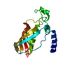
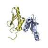
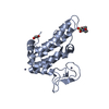
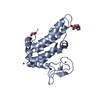
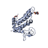
- PDB-2ucz: UBIQUITIN CONJUGATING ENZYME (UBC7) FROM SACCHAROMYCES CEREVISIAE -
+ Open data

- Basic information
| Entry | Database: PDB / ID: 2ucz | |||||||||
|---|---|---|---|---|---|---|---|---|---|---|
| Title | UBIQUITIN CONJUGATING ENZYME (UBC7) FROM SACCHAROMYCES CEREVISIAE | |||||||||
 Components Components | UBIQUITIN CONJUGATING ENZYME | |||||||||
 Keywords Keywords | UBIQUITIN CONJUGATION /  LIGASE / YEAST LIGASE / YEAST | |||||||||
| Function / homology |  Function and homology information Function and homology informationCUE1-UBC7 ubiquitin-conjugating enzyme complex / Doa10p ubiquitin ligase complex / Synthesis of active ubiquitin: roles of E1 and E2 enzymes / Hrd1p ubiquitin ligase ERAD-L complex / fungal-type cell wall organization / Antigen processing: Ubiquitination & Proteasome degradation / E2 ubiquitin-conjugating enzyme / ERAD pathway / ubiquitin conjugating enzyme activity / response to cadmium ion ...CUE1-UBC7 ubiquitin-conjugating enzyme complex / Doa10p ubiquitin ligase complex / Synthesis of active ubiquitin: roles of E1 and E2 enzymes / Hrd1p ubiquitin ligase ERAD-L complex / fungal-type cell wall organization / Antigen processing: Ubiquitination & Proteasome degradation / E2 ubiquitin-conjugating enzyme / ERAD pathway / ubiquitin conjugating enzyme activity / response to cadmium ion / ubiquitin-protein transferase activity / chromatin organization / ubiquitin-dependent protein catabolic process / protein ubiquitination / endoplasmic reticulum membrane / endoplasmic reticulum / ATP binding / cytosolSimilarity search - Function | |||||||||
| Biological species |  Saccharomyces cerevisiae (brewer's yeast) Saccharomyces cerevisiae (brewer's yeast) | |||||||||
| Method | X-RAY DIFFRACTION / MOLECULAR REPLACEMENT / Resolution: 2.93 Å | |||||||||
 Authors Authors | Cook, W.J. / Chau, V. | |||||||||
 Citation Citation | Journal: Biochemistry / Year: 1997 Title: Crystal structure of a class I ubiquitin conjugating enzyme (Ubc7) from Saccharomyces cerevisiae at 2.9 angstroms resolution. Authors: Cook, W.J. / Martin, P.D. / Edwards, B.F. / Yamazaki, R.K. / Chau, V. | |||||||||
| History |
|
- Structure visualization
| Structure viewer | Molecule: MolmilJmol/JSmol |
|---|
- Downloads & links
-Download
| PDBx/mmCIF format | 2ucz.cif.gz | 39.4 KB | Display | PDBx/mmCIF format |
|---|---|---|---|---|
| PDB format | pdb2ucz.ent.gz | 26.8 KB | Display | PDB format |
| PDBx/mmJSON format | 2ucz.json.gz | Tree view | PDBx/mmJSON format | |
| Others |  Other downloads Other downloads |
-Validation report
| Arichive directory | https://data.pdbj.org/pub/pdb/validation_reports/uc/2uczftp://data.pdbj.org/pub/pdb/validation_reports/uc/2ucz | HTTPS FTP |
|---|
-Related structure data
| Related structure data |  1aak S: Starting model for refinement |
|---|---|
| Similar structure data |


-Links
 PDBj
PDBj


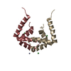
- Assembly
| Deposited unit | 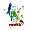
| ||||||||
|---|---|---|---|---|---|---|---|---|---|
| 1 |
| ||||||||
| Unit cell |
|
-Components
| #1: Protein | Mass: 18538.271 Da / Num. of mol.: 1 Source method: isolated from a genetically manipulated source Source: (gene. exp.) Saccharomyces cerevisiae (brewer's yeast)Cellular location: CYTOPLASM / Gene: UBC7 / Production host:  Escherichia coli (E. coli) / References: UniProt: Q02159, ubiquitin-protein ligase Escherichia coli (E. coli) / References: UniProt: Q02159, ubiquitin-protein ligase |
|---|
-Experimental details
-Experiment
| Experiment | Method: X-RAY DIFFRACTION / Number of used crystals: 2 |
|---|
- Sample preparation
| Crystal | Density Matthews: 4.36 Å3/Da / Density % sol: 72 % | ||||||||||||||||||||||||||||||
|---|---|---|---|---|---|---|---|---|---|---|---|---|---|---|---|---|---|---|---|---|---|---|---|---|---|---|---|---|---|---|---|
| Crystal grow | Method: vapor diffusion, hanging drop / pH: 7.4 Details: PROTEIN WAS CRYSTALLIZED FROM 0.6 M SODIUM CITRATE, 100 MM HEPES, PH 7.4, USING HANGING DROP TECHNIQUE AT 23 DEG, vapor diffusion - hanging drop | ||||||||||||||||||||||||||||||
| Crystal grow | *PLUS Temperature: 23 ℃ / Method: vapor diffusion | ||||||||||||||||||||||||||||||
| Components of the solutions | *PLUS
|
-Data collection
| Diffraction | Mean temperature: 295 K |
|---|---|
| Diffraction source | Source: ROTATING ANODE / Type: RIGAKU RUH3R / Wavelength: 1.5418 |
| Detector | Type: SIEMENS / Detector: AREA DETECTOR / Date: Aug 1, 1994 |
| Radiation | Monochromator: NI FILTER / Monochromatic (M) / Laue (L): M / Scattering type: x-ray |
| Radiation wavelength | Wavelength: 1.5418 Å / Relative weight: 1 |
| Reflection | Highest resolution: 2.93 Å / Num. obs: 6557 / % possible obs: 93 % / Observed criterion σ(I): -3 / Redundancy: 3.9 % / Biso Wilson estimate: 28.7 Å2 / Rsym value: 0.154 / Net I/σ(I): 8 |
| Reflection shell | Resolution: 2.93→3.11 Å / Redundancy: 2.2 % / Mean I/σ(I) obs: 1 / Rsym value: 0.327 / % possible all: 62 |
| Reflection | *PLUS Num. measured all: 25491 / Rmerge(I) obs: 0.154 |
| Reflection shell | *PLUS % possible obs: 62 % / Num. unique obs: 720 |
- Processing
| Software |
| ||||||||||||||||||||||||||||||||||||||||||||||||||||||||||||
|---|---|---|---|---|---|---|---|---|---|---|---|---|---|---|---|---|---|---|---|---|---|---|---|---|---|---|---|---|---|---|---|---|---|---|---|---|---|---|---|---|---|---|---|---|---|---|---|---|---|---|---|---|---|---|---|---|---|---|---|---|---|
| Refinement | Method to determine structure: MOLECULAR REPLACEMENT Starting model: PDB ENTRY 1AAK 1aak Resolution: 2.93→100 Å / Rfactor Rfree error: 0.014 / Data cutoff high absF: 100000 / Data cutoff low absF: 0.001 / Isotropic thermal model: OVERALL / Cross valid method: A POSTERIORI / σ(F): 0 / Details: BULK SOLVENT MODEL USED
| ||||||||||||||||||||||||||||||||||||||||||||||||||||||||||||
| Displacement parameters | Biso mean: 26.1 Å2 | ||||||||||||||||||||||||||||||||||||||||||||||||||||||||||||
| Refine analyze |
| ||||||||||||||||||||||||||||||||||||||||||||||||||||||||||||
| Refinement step | Cycle: LAST / Resolution: 2.93→100 Å
| ||||||||||||||||||||||||||||||||||||||||||||||||||||||||||||
| Refine LS restraints |
| ||||||||||||||||||||||||||||||||||||||||||||||||||||||||||||
| LS refinement shell | Resolution: 2.93→3.08 Å / Rfactor Rfree error: 0.058 / Total num. of bins used: 6
| ||||||||||||||||||||||||||||||||||||||||||||||||||||||||||||
| Xplor file |
| ||||||||||||||||||||||||||||||||||||||||||||||||||||||||||||
| Software | *PLUS Name: X-PLOR / Version: 3.851 / Classification: refinement | ||||||||||||||||||||||||||||||||||||||||||||||||||||||||||||
| Refine LS restraints | *PLUS
|
