 Movie
Movie Controller


+ Open data

- Basic information
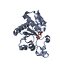
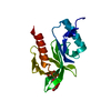
| Entry | Database: PDB / ID: 4jmk | ||||||
|---|---|---|---|---|---|---|---|
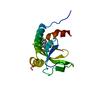
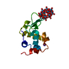
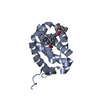
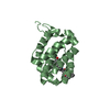
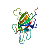
| Title | Structure of dusp8 | ||||||
 Components Components | Dual specificity protein phosphatase 8 | ||||||
 Keywords Keywords |  HYDROLASE / alpha/beta hydrolase HYDROLASE / alpha/beta hydrolase | ||||||
| Function / homology |  Function and homology information: / protein tyrosine/threonine phosphatase activity / MAP kinase tyrosine/serine/threonine phosphatase activity / peptidyl-threonine dephosphorylation / protein tyrosine/serine/threonine phosphatase activity / RAF-independent MAPK1/3 activation / mitogen-activated protein kinase binding / protein-serine/threonine phosphatase / phosphatase activity / phosphoprotein phosphatase activity ...: / protein tyrosine/threonine phosphatase activity / MAP kinase tyrosine/serine/threonine phosphatase activity / peptidyl-threonine dephosphorylation / protein tyrosine/serine/threonine phosphatase activity / RAF-independent MAPK1/3 activation / mitogen-activated protein kinase binding / protein-serine/threonine phosphatase / phosphatase activity / phosphoprotein phosphatase activity / peptidyl-tyrosine dephosphorylation / dephosphorylation / protein dephosphorylation / protein-tyrosine-phosphatase / protein tyrosine phosphatase activity / Negative regulation of MAPK pathway / nucleus / cytosol / cytoplasm Function and homology information: / protein tyrosine/threonine phosphatase activity / MAP kinase tyrosine/serine/threonine phosphatase activity / peptidyl-threonine dephosphorylation / protein tyrosine/serine/threonine phosphatase activity / RAF-independent MAPK1/3 activation / mitogen-activated protein kinase binding / protein-serine/threonine phosphatase / phosphatase activity / phosphoprotein phosphatase activity ...: / protein tyrosine/threonine phosphatase activity / MAP kinase tyrosine/serine/threonine phosphatase activity / peptidyl-threonine dephosphorylation / protein tyrosine/serine/threonine phosphatase activity / RAF-independent MAPK1/3 activation / mitogen-activated protein kinase binding / protein-serine/threonine phosphatase / phosphatase activity / phosphoprotein phosphatase activity / peptidyl-tyrosine dephosphorylation / dephosphorylation / protein dephosphorylation / protein-tyrosine-phosphatase / protein tyrosine phosphatase activity / Negative regulation of MAPK pathway / nucleus / cytosol / cytoplasmSimilarity search - Function | ||||||
| Biological species |  Homo sapiens (human) Homo sapiens (human) | ||||||
| Method | X-RAY DIFFRACTION / SYNCHROTRON / MOLECULAR REPLACEMENT / Resolution: 1.9 Å | ||||||
 Authors Authors | Jeong, D.G. / Kim, S.J. / Ryu, S.E. | ||||||
 Citation Citation | Journal: Acta Crystallogr.,Sect.D / Year: 2014 Title: The family-wide structure and function of human dual-specificity protein phosphatases. Authors: Jeong, D.G. / Wei, C.H. / Ku, B. / Jeon, T.J. / Chien, P.N. / Kim, J.K. / Park, S.Y. / Hwang, H.S. / Ryu, S.Y. / Park, H. / Kim, D.S. / Kim, S.J. / Ryu, S.E. | ||||||
| History |
|
- Structure visualization
| Structure viewer | Molecule: MolmilJmol/JSmol |
|---|
- Downloads & links
-Download
| PDBx/mmCIF format | 4jmk.cif.gz | 131.9 KB | Display | PDBx/mmCIF format |
|---|---|---|---|---|
| PDB format | pdb4jmk.ent.gz | 102.2 KB | Display | PDB format |
| PDBx/mmJSON format | 4jmk.json.gz | Tree view | PDBx/mmJSON format | |
| Others |  Other downloads Other downloads |
-Validation report
| Arichive directory | https://data.pdbj.org/pub/pdb/validation_reports/jm/4jmkftp://data.pdbj.org/pub/pdb/validation_reports/jm/4jmk | HTTPS FTP |
|---|
-Related structure data
| Related structure data |  4jmjC  4jnbC 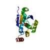 4ki9C  1zzwS C: citing same article ( S: Starting model for refinement |
|---|---|
| Similar structure data |


-Links
 PDBj
PDBj


- Assembly
| Deposited unit | 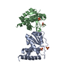
| ||||||||
|---|---|---|---|---|---|---|---|---|---|
| 1 | 
| ||||||||
| 2 | 
| ||||||||
| Unit cell |
|
-Components
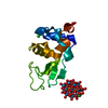
| #1: Protein | Mass: 17157.938 Da / Num. of mol.: 2 / Fragment: UNP residues 160-310 / Mutation: C246S Source method: isolated from a genetically manipulated source Source: (gene. exp.) Homo sapiens (human) / Gene: DUSP8, C11orf81, VH5 / Production host:  Escherichia coli (E. coli) Escherichia coli (E. coli)References: UniProt: Q13202, protein-serine/threonine phosphatase, protein-tyrosine-phosphatase#2: Chemical | ChemComp-SO4 / Sulfate  Mass: 96.063 Da / Num. of mol.: 4 / Source method: obtained synthetically / Formula: SO4 Mass: 96.063 Da / Num. of mol.: 4 / Source method: obtained synthetically / Formula: SO4#3: Water | ChemComp-HOH / | Water Mass: 18.015 Da / Num. of mol.: 157 / Source method: isolated from a natural source / Formula: H2O Mass: 18.015 Da / Num. of mol.: 157 / Source method: isolated from a natural source / Formula: H2O |
|---|
-Experimental details
-Experiment
| Experiment | Method: X-RAY DIFFRACTION / Number of used crystals: 1 |
|---|
- Sample preparation
| Crystal | Density Matthews: 1.97 Å3/Da / Density % sol: 37.45 % |
|---|---|
| Crystal grow | Temperature: 291 K / Method: vapor diffusion, hanging drop / pH: 7 Details: Hepes, PEG 8000, pH 7., VAPOR DIFFUSION, HANGING DROP, temperature 291K |
-Data collection
| Diffraction | Mean temperature: 100 K |
|---|---|
| Diffraction source | Source: SYNCHROTRON / Site: PAL/PLS  / Beamline: 6B / Wavelength: 1 Å / Beamline: 6B / Wavelength: 1 Å |
| Detector | Type: ADSC QUANTUM 210 / Detector: CCD / Date: Oct 4, 2006 |
| Radiation | Monochromator: Yale mirrors / Protocol: SINGLE WAVELENGTH / Monochromatic (M) / Laue (L): M / Scattering type: x-ray |
| Radiation wavelength | Wavelength: 1 Å / Relative weight: 1 |
| Reflection | Resolution: 1.9→40 Å / Num. all: 22431 / Num. obs: 22371 / % possible obs: 99.9 % / Observed criterion σ(F): 0 / Observed criterion σ(I): 0 |
| Reflection shell | Resolution: 1.9→2 Å / % possible all: 99.9 |
- Processing
| Software |
| ||||||||||||||||||||||||||||||||||||||||||||||||||||||
|---|---|---|---|---|---|---|---|---|---|---|---|---|---|---|---|---|---|---|---|---|---|---|---|---|---|---|---|---|---|---|---|---|---|---|---|---|---|---|---|---|---|---|---|---|---|---|---|---|---|---|---|---|---|---|---|
| Refinement | Method to determine structure: MOLECULAR REPLACEMENT Starting model: 1ZZW Resolution: 1.9→35.109 Å / Occupancy max: 1 / Occupancy min: 1 / SU ML: 0.19 / σ(F): 0 / Phase error: 19.8 / Stereochemistry target values: ML
| ||||||||||||||||||||||||||||||||||||||||||||||||||||||
| Solvent computation | Shrinkage radii: 0.9 Å / VDW probe radii: 1.11 Å / Solvent model: FLAT BULK SOLVENT MODEL | ||||||||||||||||||||||||||||||||||||||||||||||||||||||
| Displacement parameters | Biso max: 103.37 Å2 / Biso mean: 27.802 Å2 / Biso min: 9.7 Å2 | ||||||||||||||||||||||||||||||||||||||||||||||||||||||
| Refinement step | Cycle: LAST / Resolution: 1.9→35.109 Å
| ||||||||||||||||||||||||||||||||||||||||||||||||||||||
| Refine LS restraints |
| ||||||||||||||||||||||||||||||||||||||||||||||||||||||
| LS refinement shell | Refine-ID: X-RAY DIFFRACTION / Total num. of bins used: 8 / % reflection obs: 100 %
| ||||||||||||||||||||||||||||||||||||||||||||||||||||||
| Refinement TLS params. | Method: refined / Origin x: 14.8396 Å / Origin y: -4.8995 Å / Origin z: 4.4359 Å
| ||||||||||||||||||||||||||||||||||||||||||||||||||||||
| Refinement TLS group | Selection details: all |
