 Movie
Movie Controller

[English] 日本語
 Yorodumi
- PDB-4jlv: Crystal structure of the chimerical protein CapA1B1 in complex wi... -
+ Open data

- Basic information
| Entry | Database: PDB / ID: 4jlv | ||||||
|---|---|---|---|---|---|---|---|
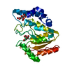
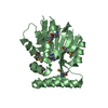
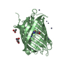
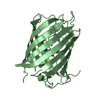
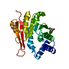
| Title | Crystal structure of the chimerical protein CapA1B1 in complex with ADP-Mg | ||||||
 Components Components | C-terminal fragment of Membrane protein CapA1, Putative uncharacterized protein capB1 | ||||||
 Keywords Keywords |  TRANSFERASE / Rossmann fold / tyrosine kinase / ATP-binding TRANSFERASE / Rossmann fold / tyrosine kinase / ATP-binding | ||||||
| Function / homology |  Function and homology information Function and homology informationcarbohydrate:proton symporter activity / polysaccharide transport / extracellular polysaccharide biosynthetic process / non-specific protein-tyrosine kinase / non-membrane spanning protein tyrosine kinase activity / protein tyrosine kinase activity / ATP binding / plasma membraneSimilarity search - Function | ||||||
| Biological species |   Staphylococcus aureus (bacteria) Staphylococcus aureus (bacteria) | ||||||
| Method | X-RAY DIFFRACTION / SYNCHROTRON / MOLECULAR REPLACEMENT / Resolution: 2.2 Å | ||||||
 Authors Authors | Gruszczyk, J. / Olivares-Illana, V. / Nourikyan, J. / Fleurie, A. / Bechet, E. / Aumont-Nicaise, M. / Gueguen-Chaignon, V. / Morera, S. / Grangeasse, C. / Nessler, S. | ||||||
 Citation Citation | Journal: Plos One / Year: 2013 Title: Comparative analysis of the Tyr-kinases CapB1 and CapB2 fused to their cognate modulators CapA1 and CapA2 from Staphylococcus aureus Authors: Gruszczyk, J. / Olivares-Illana, V. / Nourikyan, J. / Fleurie, A. / Bechet, E. / Gueguen-Chaignon, V. / Freton, C. / Aumont-Nicaise, M. / Morera, S. / Grangeasse, C. / Nessler, S. | ||||||
| History |
|
- Structure visualization
| Structure viewer | Molecule: MolmilJmol/JSmol |
|---|
- Downloads & links
-Download
| PDBx/mmCIF format | 4jlv.cif.gz | 60.5 KB | Display | PDBx/mmCIF format |
|---|---|---|---|---|
| PDB format | pdb4jlv.ent.gz | 41.9 KB | Display | PDB format |
| PDBx/mmJSON format | 4jlv.json.gz | Tree view | PDBx/mmJSON format | |
| Others |  Other downloads Other downloads |
-Validation report
| Arichive directory | https://data.pdbj.org/pub/pdb/validation_reports/jl/4jlvftp://data.pdbj.org/pub/pdb/validation_reports/jl/4jlv | HTTPS FTP |
|---|
-Related structure data
| Related structure data | 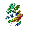 4jmpC 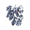 3bfvS S: Starting model for refinement C: citing same article ( |
|---|---|
| Similar structure data |


-Links
 PDBj
PDBj- Assembly
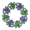
| Deposited unit | 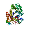
| ||||||||
|---|---|---|---|---|---|---|---|---|---|
| 1 |
| ||||||||
| Unit cell |
|
-Components
| #1: Protein | Mass: 30096.195 Da / Num. of mol.: 1 Source method: isolated from a genetically manipulated source Details: Chimera of Membrane protein CapA1 and tyrosine kinase CapB1 Source: (gene. exp.) Staphylococcus aureus (bacteria) / Gene: capA1, capB1 / Plasmid: pQE30 / Production host: Escherichia coli (E. coli) / References: UniProt: A8YPQ6, UniProt: A8YPQ8 |
|---|---|
| #2: Chemical | ChemComp-ADP / Adenosine diphosphate  Mass: 427.201 Da / Num. of mol.: 1 / Source method: obtained synthetically / Formula: C10H15N5O10P2 / Comment: ADP, energy-carrying molecule*YM Mass: 427.201 Da / Num. of mol.: 1 / Source method: obtained synthetically / Formula: C10H15N5O10P2 / Comment: ADP, energy-carrying molecule*YM |
| #3: Chemical | ChemComp-MG /   Mass: 24.305 Da / Num. of mol.: 1 / Source method: obtained synthetically / Formula: Mg Mass: 24.305 Da / Num. of mol.: 1 / Source method: obtained synthetically / Formula: Mg |
| #4: Water | ChemComp-HOH / Water Mass: 18.015 Da / Num. of mol.: 23 / Source method: isolated from a natural source / Formula: H2O Mass: 18.015 Da / Num. of mol.: 23 / Source method: isolated from a natural source / Formula: H2O |
-Experimental details
-Experiment
| Experiment | Method: X-RAY DIFFRACTION / Number of used crystals: 1 |
|---|
- Sample preparation
| Crystal | Density Matthews: 1.94 Å3/Da / Density % sol: 36.74 % |
|---|---|
| Crystal grow | Temperature: 290 K / Method: vapor diffusion / pH: 8.5 Details: 28% PEG 1000, 100mM Tris-HCl, pH 8.5, VAPOR DIFFUSION, temperature 290K |
-Data collection
| Diffraction | Mean temperature: 100 K |
|---|---|
| Diffraction source | Source: SYNCHROTRON / Site: ESRF  / Beamline: ID29 / Wavelength: 0.98 Å / Beamline: ID29 / Wavelength: 0.98 Å |
| Detector | Type: ADSC QUANTUM 210 / Detector: CCD / Date: Aug 26, 2008 |
| Radiation | Monochromator: cooled channel-cut Si(311) crystal / Protocol: SINGLE WAVELENGTH / Monochromatic (M) / Laue (L): M / Scattering type: x-ray |
| Radiation wavelength | Wavelength: 0.98 Å / Relative weight: 1 |
| Reflection | Resolution: 2.2→50 Å / Num. obs: 12422 / % possible obs: 99.7 % / Observed criterion σ(F): 0 / Observed criterion σ(I): -3 / Redundancy: 4.752 % |
| Reflection shell | Resolution: 2.2→2.33 Å / Redundancy: 4.81 % / Rmerge(I) obs: 1.113 / Mean I/σ(I) obs: 1.93 / Num. unique all: 1515 / Rsym value: 1.252 / % possible all: 99.5 |
- Processing
| Software |
| |||||||||||||||||||||||||||||||||||
|---|---|---|---|---|---|---|---|---|---|---|---|---|---|---|---|---|---|---|---|---|---|---|---|---|---|---|---|---|---|---|---|---|---|---|---|---|
| Refinement | Method to determine structure: MOLECULAR REPLACEMENT Starting model: 3BFV Resolution: 2.2→44.14 Å / Occupancy max: 1 / Occupancy min: 1 / FOM work R set: 0.7625 / SU ML: 0.25 / σ(F): 1.36 / Phase error: 29.1 / Stereochemistry target values: ML
| |||||||||||||||||||||||||||||||||||
| Solvent computation | Shrinkage radii: 0.9 Å / VDW probe radii: 1.11 Å / Solvent model: FLAT BULK SOLVENT MODEL | |||||||||||||||||||||||||||||||||||
| Displacement parameters | Biso max: 91.71 Å2 / Biso mean: 57.5635 Å2 / Biso min: 35.93 Å2 | |||||||||||||||||||||||||||||||||||
| Refinement step | Cycle: LAST / Resolution: 2.2→44.14 Å
| |||||||||||||||||||||||||||||||||||
| Refine LS restraints |
| |||||||||||||||||||||||||||||||||||
| LS refinement shell | Refine-ID: X-RAY DIFFRACTION / Total num. of bins used: 4
|
