[English] 日本語
Yorodumi
- PDB-4jkq: Crystal structure of the N-terminal region of the human ryanodine... -

+
Open data


ID or keywords:

Loading...

-
Basic information

Entry
Database: PDB / ID: 4jkq
Title
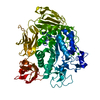
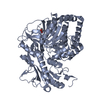
Crystal structure of the N-terminal region of the human ryanodine receptor 2
ComponentsRyanodine receptor 2
KeywordsUNKNOWN FUNCTION / beta trefoil fold
Function / homology
Function and homology information


junctional sarcoplasmic reticulum membrane / suramin binding / establishment of protein localization to endoplasmic reticulum / type B pancreatic cell apoptotic process / Purkinje myocyte to ventricular cardiac muscle cell signaling / regulation of SA node cell action potential / regulation of atrial cardiac muscle cell action potential / left ventricular cardiac muscle tissue morphogenesis / regulation of AV node cell action potential / positive regulation of ATPase-coupled calcium transmembrane transporter activity ...junctional sarcoplasmic reticulum membrane / suramin binding / establishment of protein localization to endoplasmic reticulum / type B pancreatic cell apoptotic process / Purkinje myocyte to ventricular cardiac muscle cell signaling / regulation of SA node cell action potential / regulation of atrial cardiac muscle cell action potential / left ventricular cardiac muscle tissue morphogenesis / regulation of AV node cell action potential / positive regulation of ATPase-coupled calcium transmembrane transporter activity / calcium-induced calcium release activity / sarcoplasmic reticulum calcium ion transport / ventricular cardiac muscle cell action potential / regulation of ventricular cardiac muscle cell action potential / positive regulation of sequestering of calcium ion / embryonic heart tube morphogenesis / cardiac muscle hypertrophy / ryanodine-sensitive calcium-release channel activity / response to muscle activity / release of sequestered calcium ion into cytosol by sarcoplasmic reticulum / calcium ion transport into cytosol / regulation of cardiac muscle contraction by calcium ion signaling / response to caffeine / cell communication by electrical coupling involved in cardiac conduction / response to redox state / positive regulation of heart rate / cellular response to caffeine / protein kinase A regulatory subunit binding / intracellularly gated calcium channel activity / protein kinase A catalytic subunit binding / positive regulation of the force of heart contraction / : / detection of calcium ion / smooth endoplasmic reticulum / regulation of cardiac muscle contraction / regulation of cardiac muscle contraction by regulation of the release of sequestered calcium ion / striated muscle contraction / release of sequestered calcium ion into cytosol / cardiac muscle contraction / Ion homeostasis / sarcoplasmic reticulum membrane / cellular response to epinephrine stimulus / calcium channel complex / regulation of cytosolic calcium ion concentration / response to muscle stretch / regulation of heart rate / sarcoplasmic reticulum / establishment of localization in cell / calcium-mediated signaling / calcium channel activity / Stimuli-sensing channels / sarcolemma / Z disc / intracellular calcium ion homeostasis / calcium ion transport / : / transmembrane transporter binding / calmodulin binding / response to hypoxia / calcium ion binding / enzyme binding / protein-containing complex / membrane / identical protein binding / plasma membrane
Similarity search - Function
IP3 receptor type 1 binding core, RIH domain / : / Ryanodine receptor junctional solenoid repeat / Ryanodine receptor, SPRY domain 2 / Ryanodine Receptor TM 4-6 / Ryanodine receptor / Ryanodine receptor, SPRY domain 1 / Ryanodine receptor, SPRY domain 3 / Ryanodine Receptor TM 4-6 / Ryanodine receptor Ryr ...IP3 receptor type 1 binding core, RIH domain / : / Ryanodine receptor junctional solenoid repeat / Ryanodine receptor, SPRY domain 2 / Ryanodine Receptor TM 4-6 / Ryanodine receptor / Ryanodine receptor, SPRY domain 1 / Ryanodine receptor, SPRY domain 3 / Ryanodine Receptor TM 4-6 / Ryanodine receptor Ryr / RyR domain / RyR/IP3 receptor binding core, RIH domain superfamily / : / RyR/IP3R Homology associated domain / Inositol 1,4,5-trisphosphate/ryanodine receptor / RIH domain / RyR and IP3R Homology associated / Inositol 1,4,5-trisphosphate/ryanodine receptor / RIH domain / MIR motif / MIR domain / MIR domain profile. / Domain in ryanodine and inositol trisphosphate receptors and protein O-mannosyltransferases / Mir domain superfamily / SPRY domain / B30.2/SPRY domain / B30.2/SPRY domain profile. / Leucine-rich Repeat Variant / SPRY domain / B30.2/SPRY domain superfamily / Domain in SPla and the RYanodine Receptor. / Trefoil (Acidic Fibroblast Growth Factor, subunit A) - #50 / Trefoil (Acidic Fibroblast Growth Factor, subunit A) / Trefoil / EF-hand domain pair / EF-hand domain / Ion transport domain / Ion transport protein / Alpha Horseshoe / EF-hand domain pair / Concanavalin A-like lectin/glucanase domain superfamily / Mainly Beta / Mainly Alpha
Similarity search - Domain/homology
Ryanodine receptor 2
Similarity search - Component
Biological speciesHomo sapiens (human)
MethodX-RAY DIFFRACTION / SYNCHROTRON / MOLECULAR REPLACEMENT / Resolution: 2.39 Å
AuthorsBauerova, V. / Sevcik, J.
CitationJournal: Acta Crystallogr.,Sect.D / Year: 2014
Title: Structural insights into the human RyR2 N-terminal region involved in cardiac arrhythmias.
Authors: Borko, L. / Bauerova-Hlinkova, V. / Hostinova, E. / Gasperik, J. / Beck, K. / Lai, F.A. / Zahradnikova, A. / Sevcik, J.
History
DepositionMar 11, 2013Deposition site: RCSB / Processing site: RCSB
Revision 1.0Apr 2, 2014Provider: repository / Type: Initial release
Revision 1.1Nov 12, 2014Group: Database references
Revision 1.2Dec 24, 2014Group: Database references
Revision 1.3Sep 20, 2023Group: Data collection / Database references / Refinement description
Category: chem_comp_atom / chem_comp_bond ...chem_comp_atom / chem_comp_bond / database_2 / pdbx_initial_refinement_model
Item: _database_2.pdbx_DOI / _database_2.pdbx_database_accession

-
Structure visualization

Structure viewerMolecule:
MolmilJmol/JSmol

Downloads & links

-
Assembly

Deposited unit
A: Ryanodine receptor 2


Theoretical massNumber of molelcules
Total (without water)67,9171
Polymers67,9171
Non-polymers00
Water1,42379
1


  • Idetical with deposited unit
  • defined by author&software
TypeNameSymmetry operationNumber
identity operation1_555x,y,z1
MethodPISA
Unit cell
Length a, b, c (Å)75.450, 75.450, 248.840
Angle α, β, γ (deg.)90.00, 90.00, 90.00
Int Tables number94
Space group name H-MP42212

-
Components

#1: Protein Ryanodine receptor 2 / / RYR-2 / RyR2 / hRYR-2 / Cardiac muscle ryanodine receptor / Cardiac muscle ryanodine receptor- ...RYR-2 / RyR2 / hRYR-2 / Cardiac muscle ryanodine receptor / Cardiac muscle ryanodine receptor-calcium release channel / Type 2 ryanodine receptor


Mass: 67916.977 Da / Num. of mol.: 1 / Fragment: N-terminal region
Source method: isolated from a genetically manipulated source
Source: (gene. exp.) Homo sapiens (human) / Gene: RYR2 / Plasmid: pET28a / Production host: Escherichia coli (E. coli) / Strain (production host): BL21 / References: UniProt: Q92736
#2: Water ChemComp-HOH / water / Water


Mass: 18.015 Da / Num. of mol.: 79 / Source method: isolated from a natural source / Formula: H2O

-
Experimental details

-
Experiment

ExperimentMethod: X-RAY DIFFRACTION / Number of used crystals: 1

-
Sample preparation

CrystalDensity Matthews: 2.59 Å3/Da / Density % sol: 52.52 %
Crystal growTemperature: 295 K / Method: vapor diffusion, hanging drop / pH: 8
Details: 20 mM TRIS-HCl, 150 mM NaCl, 10% glycerol, 7mM mercaptoethanol, 1 mM CHAPS, 0.1% betaine, 100 mM HEPES, 200 mM ammonium formate, 21% PEG 3350, pH 8.0, VAPOR DIFFUSION, HANGING DROP, temperature 295K

-
Data collection

DiffractionMean temperature: 100 K
Diffraction sourceSource: SYNCHROTRON / Site: BESSY / Beamline: 14.1 / Wavelength: 0.918 Å
DetectorType: MARMOSAIC 225 mm CCD / Detector: CCD / Date: Oct 5, 2012
RadiationMonochromator: MIRRORS / Protocol: SINGLE WAVELENGTH / Monochromatic (M) / Laue (L): M / Scattering type: x-ray
Radiation wavelengthWavelength: 0.918 Å / Relative weight: 1
ReflectionResolution: 2.39→124.42 Å / Num. obs: 29469 / % possible obs: 100 % / Observed criterion σ(F): 2 / Observed criterion σ(I): 1 / Redundancy: 13.8 % / Rmerge(I) obs: 0.16 / Rsym value: 0.15 / Net I/σ(I): 12.2
Reflection shellResolution: 2.39→2.52 Å / % possible all: 95

-
Processing

Software
NameVersionClassification
MAR345dtbdata collection
MOLREPphasing
REFMAC5.6.0117refinement
MOSFLMdata reduction
SCALAdata scaling
RefinementMethod to determine structure: MOLECULAR REPLACEMENT
Starting model: PDB entry 2XOA
Resolution: 2.39→44.77 Å / Cor.coef. Fo:Fc: 0.94 / Cor.coef. Fo:Fc free: 0.912 / SU B: 18.374 / SU ML: 0.203 / Cross valid method: THROUGHOUT / ESU R: 0.316 / ESU R Free: 0.24 / Stereochemistry target values: MAXIMUM LIKELIHOOD / Details: HYDROGENS HAVE BEEN USED IF PRESENT IN THE INPUT
RfactorNum. reflection% reflectionSelection details
Rfree0.26087 1444 4.9 %RANDOM
Rwork0.22405 ---
obs0.2258 27960 99.35 %-
Solvent computationIon probe radii: 0.8 Å / Shrinkage radii: 0.8 Å / VDW probe radii: 1.2 Å / Solvent model: MASK
Displacement parametersBiso mean: 62.542 Å2
Baniso -1Baniso -2Baniso -3
1-1.84 Å2-0 Å20 Å2
2--1.84 Å20 Å2
3----3.68 Å2
Refinement stepCycle: LAST / Resolution: 2.39→44.77 Å
ProteinNucleic acidLigandSolventTotal
Num. atoms3934 0 0 79 4013
Refine LS restraints
Refine-IDTypeDev idealDev ideal targetNumber
X-RAY DIFFRACTIONr_bond_refined_d0.0080.0194009
X-RAY DIFFRACTIONr_bond_other_d
X-RAY DIFFRACTIONr_angle_refined_deg1.1851.9465411
X-RAY DIFFRACTIONr_angle_other_deg
X-RAY DIFFRACTIONr_dihedral_angle_1_deg5.9085496
X-RAY DIFFRACTIONr_dihedral_angle_2_deg37.94123.316190
X-RAY DIFFRACTIONr_dihedral_angle_3_deg15.95915708
X-RAY DIFFRACTIONr_dihedral_angle_4_deg15.9431533
X-RAY DIFFRACTIONr_chiral_restr0.0820.2602
X-RAY DIFFRACTIONr_gen_planes_refined0.0040.023010
X-RAY DIFFRACTIONr_gen_planes_other
X-RAY DIFFRACTIONr_nbd_refined
X-RAY DIFFRACTIONr_nbd_other
X-RAY DIFFRACTIONr_nbtor_refined
X-RAY DIFFRACTIONr_nbtor_other
X-RAY DIFFRACTIONr_xyhbond_nbd_refined
X-RAY DIFFRACTIONr_xyhbond_nbd_other
X-RAY DIFFRACTIONr_metal_ion_refined
X-RAY DIFFRACTIONr_metal_ion_other
X-RAY DIFFRACTIONr_symmetry_vdw_refined
X-RAY DIFFRACTIONr_symmetry_vdw_other
X-RAY DIFFRACTIONr_symmetry_hbond_refined
X-RAY DIFFRACTIONr_symmetry_hbond_other
X-RAY DIFFRACTIONr_symmetry_metal_ion_refined
X-RAY DIFFRACTIONr_symmetry_metal_ion_other
X-RAY DIFFRACTIONr_mcbond_it
X-RAY DIFFRACTIONr_mcbond_other
X-RAY DIFFRACTIONr_mcangle_it
X-RAY DIFFRACTIONr_mcangle_other
X-RAY DIFFRACTIONr_scbond_it
X-RAY DIFFRACTIONr_scbond_other
X-RAY DIFFRACTIONr_scangle_it
X-RAY DIFFRACTIONr_scangle_other
X-RAY DIFFRACTIONr_long_range_B_refined
X-RAY DIFFRACTIONr_long_range_B_other
X-RAY DIFFRACTIONr_rigid_bond_restr3.88633986
X-RAY DIFFRACTIONr_sphericity_free74.934534
X-RAY DIFFRACTIONr_sphericity_bonded26.95653927
LS refinement shellResolution: 2.39→2.449 Å / Total num. of bins used: 20
RfactorNum. reflection% reflection
Rfree0.334 74 -
Rwork0.336 1598 -
obs--90.04 %
Refinement TLS params.

Method: refined / Refine-ID: X-RAY DIFFRACTION

IDL112)L122)L132)L222)L232)L332)S11 (Å °)S12 (Å °)S13 (Å °)S21 (Å °)S22 (Å °)S23 (Å °)S31 (Å °)S32 (Å °)S33 (Å °)T112)T122)T132)T222)T232)T332)Origin x (Å)Origin y (Å)Origin z (Å)
11.910.18960.05351.01970.11224.35550.0572-0.0460.0118-0.02850.00620.0254-0.1208-0.0735-0.06340.23560.06850.0560.135-0.06660.130520.411-32.984-49.423
23.1634-0.7673-1.09582.8850.47185.4209-0.2331-0.43060.13340.30510.2637-0.1270.13890.6109-0.03060.40380.2465-0.02980.2238-0.02820.009422.385-25.446-17.451
30.88071.168-0.72612.3923-1.34952.96270.0821-0.06090.1038-0.0320.0160.20.19270.3262-0.09810.20790.10320.01360.13050.00580.14278.131-7.224-38.918
Refinement TLS group
IDRefine-IDRefine TLS-IDAuth asym-IDAuth seq-ID
1X-RAY DIFFRACTION1A11 - 223
2X-RAY DIFFRACTION2A224 - 408
3X-RAY DIFFRACTION3A409 - 544

+
About Yorodumi

-
News

-
Feb 9, 2022. New format data for meta-information of EMDB entries

New format data for meta-information of EMDB entries

  • Version 3 of the EMDB header file is now the official format.
  • The previous official version 1.9 will be removed from the archive.

Related info.:EMDB header

External links:wwPDB to switch to version 3 of the EMDB data model

-
Aug 12, 2020. Covid-19 info

Covid-19 info

URL: https://pdbj.org/emnavi/covid19.php

New page: Covid-19 featured information page in EM Navigator.

Related info.:Covid-19 info / Mar 5, 2020. Novel coronavirus structure data

+
Mar 5, 2020. Novel coronavirus structure data

Novel coronavirus structure data

Related info.:Yorodumi Speices / Aug 12, 2020. Covid-19 info

External links:COVID-19 featured content - PDBj / Molecule of the Month (242):Coronavirus Proteases

+
Jan 31, 2019. EMDB accession codes are about to change! (news from PDBe EMDB page)

EMDB accession codes are about to change! (news from PDBe EMDB page)

  • The allocation of 4 digits for EMDB accession codes will soon come to an end. Whilst these codes will remain in use, new EMDB accession codes will include an additional digit and will expand incrementally as the available range of codes is exhausted. The current 4-digit format prefixed with “EMD-” (i.e. EMD-XXXX) will advance to a 5-digit format (i.e. EMD-XXXXX), and so on. It is currently estimated that the 4-digit codes will be depleted around Spring 2019, at which point the 5-digit format will come into force.
  • The EM Navigator/Yorodumi systems omit the EMD- prefix.

Related info.:Q: What is EMD? / ID/Accession-code notation in Yorodumi/EM Navigator

External links:EMDB Accession Codes are Changing Soon! / Contact to PDBj

+
Jul 12, 2017. Major update of PDB

Major update of PDB

  • wwPDB released updated PDB data conforming to the new PDBx/mmCIF dictionary.
  • This is a major update changing the version number from 4 to 5, and with Remediation, in which all the entries are updated.
  • In this update, many items about electron microscopy experimental information are reorganized (e.g. em_software).
  • Now, EM Navigator and Yorodumi are based on the updated data.

External links:wwPDB Remediation / Enriched Model Files Conforming to OneDep Data Standards Now Available in the PDB FTP Archive

-
Yorodumi

Thousand views of thousand structures

  • Yorodumi is a browser for structure data from EMDB, PDB, SASBDB, etc.
  • This page is also the successor to EM Navigator detail page, and also detail information page/front-end page for Omokage search.
  • The word "yorodu" (or yorozu) is an old Japanese word meaning "ten thousand". "mi" (miru) is to see.

Related info.:EMDB / PDB / SASBDB / Comparison of 3 databanks / Yorodumi Search / Aug 31, 2016. New EM Navigator & Yorodumi / Yorodumi Papers / Jmol/JSmol / Function and homology information / Changes in new EM Navigator and Yorodumi

Read more