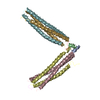
 Movie
Movie Controller


+ Open data

- Basic information
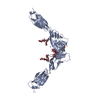
| Entry | Database: PDB / ID: 4jjy | ||||||
|---|---|---|---|---|---|---|---|
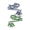
| Title | Alix V domain | ||||||
 Components Components | Programmed cell death 6-interacting protein | ||||||
 Keywords Keywords |  PROTEIN BINDING / Ubiquitin / Endosome / Membrane Trafficking / virus budding / ESCRTI PROTEIN BINDING / Ubiquitin / Endosome / Membrane Trafficking / virus budding / ESCRTI | ||||||
| Function / homology |  Function and homology information Function and homology informationproteinase activated receptor binding / actomyosin contractile ring assembly / ubiquitin-independent protein catabolic process via the multivesicular body sorting pathway / regulation of extracellular exosome assembly / viral budding / extracellular exosome biogenesis / maintenance of epithelial cell apical/basal polarity / positive regulation of extracellular exosome assembly / regulation of membrane permeability / regulation of centrosome duplication ...proteinase activated receptor binding / actomyosin contractile ring assembly / ubiquitin-independent protein catabolic process via the multivesicular body sorting pathway / regulation of extracellular exosome assembly / viral budding / extracellular exosome biogenesis / maintenance of epithelial cell apical/basal polarity / positive regulation of extracellular exosome assembly / regulation of membrane permeability / regulation of centrosome duplication / midbody abscission / multivesicular body sorting pathway / bicellular tight junction assembly / actomyosin / positive regulation of exosomal secretion / multivesicular body assembly / Flemming body / RIPK1-mediated regulated necrosis / viral budding via host ESCRT complex / mitotic cytokinesis / Uptake and function of anthrax toxins / immunological synapse / bicellular tight junction / endoplasmic reticulum exit site / macroautophagy / Budding and maturation of HIV virion / protein homooligomerization / Regulation of necroptotic cell death / calcium-dependent protein binding / extracellular vesicle / melanosome / protein transport / endosome / focal adhesion / centrosome / apoptotic process / protein homodimerization activity / extracellular exosome / membrane / cytosolSimilarity search - Function | ||||||
| Biological species |  Homo sapiens (human) Homo sapiens (human) | ||||||
| Method | X-RAY DIFFRACTION / SYNCHROTRON / SAD / Resolution: 6.503 Å | ||||||
 Authors Authors | Pashkova, N. / Gakhar, L. / Yu, L. / Piper, R.C. | ||||||
 Citation Citation | Journal: Dev.Cell / Year: 2013 Title: The yeast alix homolog bro1 functions as a ubiquitin receptor for protein sorting into multivesicular endosomes. Authors: Pashkova, N. / Gakhar, L. / Winistorfer, S.C. / Sunshine, A.B. / Rich, M. / Dunham, M.J. / Yu, L. / Piper, R.C. | ||||||
| History |
|
- Structure visualization
| Structure viewer | Molecule: MolmilJmol/JSmol |
|---|
- Downloads & links
-Download
| PDBx/mmCIF format | 4jjy.cif.gz | 125.7 KB | Display | PDBx/mmCIF format |
|---|---|---|---|---|
| PDB format | pdb4jjy.ent.gz | 105.3 KB | Display | PDB format |
| PDBx/mmJSON format | 4jjy.json.gz | Tree view | PDBx/mmJSON format | |
| Others |  Other downloads Other downloads |
-Validation report
| Arichive directory | https://data.pdbj.org/pub/pdb/validation_reports/jj/4jjyftp://data.pdbj.org/pub/pdb/validation_reports/jj/4jjy | HTTPS FTP |
|---|
-Related structure data


-Links
 PDBj
PDBj


- Assembly
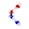
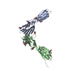
| Deposited unit | 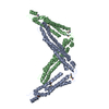
| ||||||||
|---|---|---|---|---|---|---|---|---|---|
| 1 | 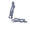
| ||||||||
| 2 | 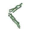
| ||||||||
| Unit cell |
|
-Components
| #1: Protein | Mass: 40898.375 Da / Num. of mol.: 2 Source method: isolated from a genetically manipulated source Source: (gene. exp.) Homo sapiens (human) / Gene: PDCD6IP, AIP1, ALIX, KIAA1375 / Production host:  Escherichia coli (E. coli) / References: UniProt: Q8WUM4 Escherichia coli (E. coli) / References: UniProt: Q8WUM4 |
|---|
-Experimental details
-Experiment
| Experiment | Method: X-RAY DIFFRACTION / Number of used crystals: 1 |
|---|
- Sample preparation
| Crystal | Density Matthews: 5.71 Å3/Da / Density % sol: 78.45 % |
|---|---|
| Crystal grow | Temperature: 277 K / Method: vapor diffusion, hanging drop / pH: 5 Details: 0.1 M sodium citrate pH 5, 6-8% PEG 8000, 5-10% ethylene glycol, VAPOR DIFFUSION, HANGING DROP, temperature 277K |
-Data collection
| Diffraction | Mean temperature: 100 K |
|---|---|
| Diffraction source | Source: SYNCHROTRON / Site: ALS  / Beamline: 4.2.2 / Wavelength: 0.976 Å / Beamline: 4.2.2 / Wavelength: 0.976 Å |
| Detector | Type: NOIR-1 / Detector: CCD / Date: Nov 30, 2011 |
| Radiation | Monochromator: Rosenbaum-Rock monochromator double crystal focussing Protocol: SINGLE WAVELENGTH / Monochromatic (M) / Laue (L): M / Scattering type: x-ray |
| Radiation wavelength | Wavelength: 0.976 Å / Relative weight: 1 |
| Reflection | Resolution: 6.5→39.56 Å / Num. all: 3774 / Num. obs: 3774 / % possible obs: 100 % / Observed criterion σ(I): 5 / Redundancy: 21.41 % / Biso Wilson estimate: 468.9 Å2 / Rmerge(I) obs: 0.061 / Net I/σ(I): 23.3 |
| Reflection shell | Resolution: 6.5→6.73 Å / Redundancy: 22.27 % / Rmerge(I) obs: 0.748 / Mean I/σ(I) obs: 2.1 / Num. unique all: 370 / % possible all: 100 |
- Processing
| Software |
| ||||||||||||||||||||||||||||
|---|---|---|---|---|---|---|---|---|---|---|---|---|---|---|---|---|---|---|---|---|---|---|---|---|---|---|---|---|---|
| Refinement | Method to determine structure: SAD / Resolution: 6.503→39.557 Å / SU ML: 1.09 / σ(F): 0 / Phase error: 33.4 / Stereochemistry target values: MLHL
| ||||||||||||||||||||||||||||
| Solvent computation | Shrinkage radii: 0.9 Å / VDW probe radii: 1.11 Å / Solvent model: FLAT BULK SOLVENT MODEL | ||||||||||||||||||||||||||||
| Refinement step | Cycle: LAST / Resolution: 6.503→39.557 Å
| ||||||||||||||||||||||||||||
| Refine LS restraints |
| ||||||||||||||||||||||||||||
| LS refinement shell |
|
