 Movie
Movie Controller

[English] 日本語
 Yorodumi
- PDB-4bfi: Structure of the complex of the extracellular portions of mouse C... -
+ Open data

- Basic information
| Entry | Database: PDB / ID: 4bfi | ||||||
|---|---|---|---|---|---|---|---|
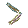
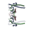
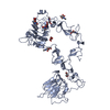
| Title | Structure of the complex of the extracellular portions of mouse CD200R and mouse CD200 | ||||||
 Components Components |
| ||||||
 Keywords Keywords |  IMMUNE SYSTEM / PAIRED RECEPTOR / IG DOMAINS / VIRAL MIMICRY / LEUKAEMIA MIMICRY / LEUKAEMIA IMMUNE SYSTEM / PAIRED RECEPTOR / IG DOMAINS / VIRAL MIMICRY / LEUKAEMIA MIMICRY / LEUKAEMIA | ||||||
| Function / homology |  Function and homology information Function and homology informationpositive regulation of arginase activity / positive regulation of protein-glutamine gamma-glutamyltransferase activity / negative regulation of T cell migration / negative regulation of matrix metallopeptidase secretion / negative regulation of macrophage migration / : / positive regulation of CREB transcription factor activity / protein binding involved in heterotypic cell-cell adhesion / negative regulation of macrophage activation / negative regulation of neuroinflammatory response ...positive regulation of arginase activity / positive regulation of protein-glutamine gamma-glutamyltransferase activity / negative regulation of T cell migration / negative regulation of matrix metallopeptidase secretion / negative regulation of macrophage migration / : / positive regulation of CREB transcription factor activity / protein binding involved in heterotypic cell-cell adhesion / negative regulation of macrophage activation / negative regulation of neuroinflammatory response / positive regulation of transforming growth factor beta production / regulation of neuroinflammatory response / heterotypic cell-cell adhesion / Immunoregulatory interactions between a Lymphoid and a non-Lymphoid cell / negative regulation of NF-kappaB transcription factor activity / negative regulation of interleukin-6 production / regulation of immune response / cell-cell adhesion / signaling receptor activity / cell body / membrane => GO:0016020 / receptor complex / neuron projection / negative regulation of cell population proliferation / external side of plasma membrane / axon / neuronal cell body / cell surface / plasma membraneSimilarity search - Function | ||||||
| Biological species |  MUS MUSCULUS (house mouse) MUS MUSCULUS (house mouse) | ||||||
| Method | X-RAY DIFFRACTION / SYNCHROTRON / MOLECULAR REPLACEMENT / Resolution: 3.22 Å | ||||||
 Authors Authors | Hatherley, D. / Lea, S.M. / Johnson, S. / Barclay, A.N. | ||||||
 Citation Citation | Journal: Structure / Year: 2013 Title: Structures of Cd200/Cd200 Receptor Family and Implications for Topology, Regulation, and Evolution Authors: Hatherley, D. / Lea, S.M. / Johnson, S. / Barclay, A.N. | ||||||
| History |
|
- Structure visualization
| Structure viewer | Molecule: MolmilJmol/JSmol |
|---|
- Downloads & links
-Download
| PDBx/mmCIF format | 4bfi.cif.gz | 95.7 KB | Display | PDBx/mmCIF format |
|---|---|---|---|---|
| PDB format | pdb4bfi.ent.gz | 73.1 KB | Display | PDB format |
| PDBx/mmJSON format | 4bfi.json.gz | Tree view | PDBx/mmJSON format | |
| Others |  Other downloads Other downloads |
-Validation report
| Arichive directory | https://data.pdbj.org/pub/pdb/validation_reports/bf/4bfiftp://data.pdbj.org/pub/pdb/validation_reports/bf/4bfi | HTTPS FTP |
|---|
-Related structure data
| Related structure data | 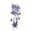 4bfeC  4bfgSC C: citing same article ( S: Starting model for refinement |
|---|---|
| Similar structure data |


-Links
 PDBj
PDBj


- Assembly
| Deposited unit | 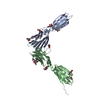
| ||||||||
|---|---|---|---|---|---|---|---|---|---|
| 1 |
| ||||||||
| Unit cell |
|
-Components
-Protein / Antibody / Sugars , 3 types, 13 molecules AB
| #1: Protein | Mass: 23586.258 Da / Num. of mol.: 1 / Fragment: EXTRACELLULAR DOMAIN, RESIDUES 26-228 Source method: isolated from a genetically manipulated source Source: (gene. exp.) MUS MUSCULUS (house mouse) / Plasmid: PEE14 / Cell line (production host): CHO / Production host:  CRICETULUS GRISEUS (Chinese hamster) / Variant (production host): LEC3.2.8.1 / References: UniProt: Q9ES57 CRICETULUS GRISEUS (Chinese hamster) / Variant (production host): LEC3.2.8.1 / References: UniProt: Q9ES57 |
|---|---|
| #2: Antibody | Mass: 23718.795 Da / Num. of mol.: 1 / Fragment: EXTRACELLULAR DOMAIN, RESIDUES 31-232 Source method: isolated from a genetically manipulated source Source: (gene. exp.) MUS MUSCULUS (house mouse) / Plasmid: PEE14 / Cell line (production host): CHO / Production host: CRICETULUS GRISEUS (Chinese hamster) / Variant (production host): LEC3.2.8.1 / References: UniProt: O54901 |
| #3: Sugar | ChemComp-NAG / N-Acetylglucosamine Type: D-saccharide, beta linking / Mass: 221.208 Da / Num. of mol.: 11 Type: D-saccharide, beta linking / Mass: 221.208 Da / Num. of mol.: 11Source method: isolated from a genetically manipulated source Formula: C8H15NO6 |
-Non-polymers , 3 types, 29 molecules 


| #4: Chemical | ChemComp-EDO / Ethylene glycol Mass: 62.068 Da / Num. of mol.: 1 / Source method: obtained synthetically / Formula: C2H6O2 Mass: 62.068 Da / Num. of mol.: 1 / Source method: obtained synthetically / Formula: C2H6O2 |
|---|---|
| #5: Chemical | ChemComp-CYS / Cysteine Type: L-peptide linking / Mass: 121.158 Da / Num. of mol.: 1 / Source method: obtained synthetically / Formula: C3H7NO2S Type: L-peptide linking / Mass: 121.158 Da / Num. of mol.: 1 / Source method: obtained synthetically / Formula: C3H7NO2S |
| #6: Water | ChemComp-HOH / WaterMass: 18.015 Da / Num. of mol.: 27 / Source method: isolated from a natural source / Formula: H2O |
-Details
| Sequence details | NUMBERING IN THE PDB IS BASED ON THE MATURE SEQUENCE AS DETERMINED |
|---|
-Experimental details
-Experiment
| Experiment | Method: X-RAY DIFFRACTION / Number of used crystals: 1 |
|---|
- Sample preparation
| Crystal | Density Matthews: 4.48 Å3/Da / Density % sol: 73 % / Description: NONE |
|---|---|
| Crystal grow | pH: 7 / Details: 1.0M IMIDAZOLE PH7.0 |
-Data collection
| Diffraction | Mean temperature: 100 K |
|---|---|
| Diffraction source | Source: SYNCHROTRON / Site: Diamond  / Beamline: I04 / Wavelength: 0.984 / Beamline: I04 / Wavelength: 0.984 |
| Detector | Type: ADSC CCD / Detector: CCD / Date: May 21, 2012 |
| Radiation | Protocol: SINGLE WAVELENGTH / Monochromatic (M) / Laue (L): M / Scattering type: x-ray |
| Radiation wavelength | Wavelength: 0.984 Å / Relative weight: 1 |
| Reflection | Resolution: 3.22→86.04 Å / Num. obs: 16034 / % possible obs: 99.6 % / Observed criterion σ(I): 2 / Redundancy: 6.3 % / Rmerge(I) obs: 0.12 / Net I/σ(I): 10.2 |
| Reflection shell | Resolution: 3.22→3.31 Å / Redundancy: 6.6 % / Rmerge(I) obs: 0.68 / Mean I/σ(I) obs: 2.9 / % possible all: 99.5 |
- Processing
| Software |
| ||||||||||||||||||||||||||||||||||||||||||||||||||||||||||||||||||||||||||||||||||||||||||||||||||||||||||||||||||||||||||||||||||||||||||||||||||||||||||||||||||||||||||||||||||||||
|---|---|---|---|---|---|---|---|---|---|---|---|---|---|---|---|---|---|---|---|---|---|---|---|---|---|---|---|---|---|---|---|---|---|---|---|---|---|---|---|---|---|---|---|---|---|---|---|---|---|---|---|---|---|---|---|---|---|---|---|---|---|---|---|---|---|---|---|---|---|---|---|---|---|---|---|---|---|---|---|---|---|---|---|---|---|---|---|---|---|---|---|---|---|---|---|---|---|---|---|---|---|---|---|---|---|---|---|---|---|---|---|---|---|---|---|---|---|---|---|---|---|---|---|---|---|---|---|---|---|---|---|---|---|---|---|---|---|---|---|---|---|---|---|---|---|---|---|---|---|---|---|---|---|---|---|---|---|---|---|---|---|---|---|---|---|---|---|---|---|---|---|---|---|---|---|---|---|---|---|---|---|---|---|
| Refinement | Method to determine structure: MOLECULAR REPLACEMENT Starting model: PDB ENTRY 4BFG Resolution: 3.22→86.04 Å / Cor.coef. Fo:Fc: 0.937 / Cor.coef. Fo:Fc free: 0.917 / SU B: 17.879 / SU ML: 0.285 / Cross valid method: THROUGHOUT / ESU R: 0.756 / ESU R Free: 0.378 / Stereochemistry target values: MAXIMUM LIKELIHOOD / Details: HYDROGENS HAVE BEEN ADDED IN THE RIDING POSITIONS.
| ||||||||||||||||||||||||||||||||||||||||||||||||||||||||||||||||||||||||||||||||||||||||||||||||||||||||||||||||||||||||||||||||||||||||||||||||||||||||||||||||||||||||||||||||||||||
| Solvent computation | Ion probe radii: 0.8 Å / Shrinkage radii: 0.8 Å / VDW probe radii: 1.2 Å / Solvent model: MASK | ||||||||||||||||||||||||||||||||||||||||||||||||||||||||||||||||||||||||||||||||||||||||||||||||||||||||||||||||||||||||||||||||||||||||||||||||||||||||||||||||||||||||||||||||||||||
| Displacement parameters | Biso mean: 94.502 Å2
| ||||||||||||||||||||||||||||||||||||||||||||||||||||||||||||||||||||||||||||||||||||||||||||||||||||||||||||||||||||||||||||||||||||||||||||||||||||||||||||||||||||||||||||||||||||||
| Refinement step | Cycle: LAST / Resolution: 3.22→86.04 Å
| ||||||||||||||||||||||||||||||||||||||||||||||||||||||||||||||||||||||||||||||||||||||||||||||||||||||||||||||||||||||||||||||||||||||||||||||||||||||||||||||||||||||||||||||||||||||
| Refine LS restraints |
|