 Movie
Movie Controller

[English] 日本語
 Yorodumi
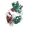
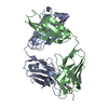
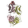
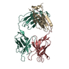
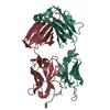
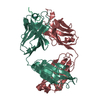
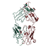
- PDB-4jj5: CRYSTAL STRUCTURE OF THE Fab FRAGMENT OF 1C2, A MONOCLONAL ANTIBO... -
+ Open data

- Basic information
| Entry | Database: PDB / ID: 4jj5 | ||||||
|---|---|---|---|---|---|---|---|
| Title | CRYSTAL STRUCTURE OF THE Fab FRAGMENT OF 1C2, A MONOCLONAL ANTIBODY SPECIFIC for POLY-GLUTAMINE | ||||||
 Components Components |
| ||||||
 Keywords Keywords |  IMMUNE SYSTEM / 1C2 / Vlx / Neurodegeneration / Polyglutamine Disease / Amyloid Disease / Immunoglobulin fold / Polyglutamine IMMUNE SYSTEM / 1C2 / Vlx / Neurodegeneration / Polyglutamine Disease / Amyloid Disease / Immunoglobulin fold / Polyglutamine | ||||||
| Function / homology | Immunoglobulins / Immunoglobulin-like / Sandwich / Mainly Beta Function and homology information Function and homology information | ||||||
| Biological species |  Mus musculus (house mouse) Mus musculus (house mouse) | ||||||
| Method | X-RAY DIFFRACTION / SYNCHROTRON / MOLECULAR REPLACEMENT / Resolution: 2.445 Å | ||||||
 Authors Authors | Klein, F.A.C. / Zeder-Lutz, G. / Cousido-Siah, A. / Mitschler, A. / Katz, A. / Eberling, P. / Mandel, J.L. / Podjarny, A. / Trottier, Y. | ||||||
 Citation Citation | Journal: Hum.Mol.Genet. / Year: 2013 Title: Linear and extended: a common polyglutamine conformation recognized by the three antibodies MW1, 1C2 and 3B5H10. Authors: Klein, F.A. / Zeder-Lutz, G. / Cousido-Siah, A. / Mitschler, A. / Katz, A. / Eberling, P. / Mandel, J.L. / Podjarny, A. / Trottier, Y. | ||||||
| History |
|
- Structure visualization
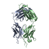
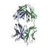
| Structure viewer | Molecule: MolmilJmol/JSmol |
|---|
- Downloads & links
-Download
| PDBx/mmCIF format | 4jj5.cif.gz | 89.4 KB | Display | PDBx/mmCIF format |
|---|---|---|---|---|
| PDB format | pdb4jj5.ent.gz | 67.4 KB | Display | PDB format |
| PDBx/mmJSON format | 4jj5.json.gz | Tree view | PDBx/mmJSON format | |
| Others |  Other downloads Other downloads |
-Validation report
| Arichive directory | https://data.pdbj.org/pub/pdb/validation_reports/jj/4jj5ftp://data.pdbj.org/pub/pdb/validation_reports/jj/4jj5 | HTTPS FTP |
|---|
-Related structure data
| Related structure data | 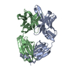 4isvSC S: Starting model for refinement C: citing same article ( |
|---|---|
| Similar structure data |
-Links
 PDBj
PDBj

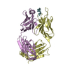
- Assembly
| Deposited unit | 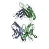
| |||||||||
|---|---|---|---|---|---|---|---|---|---|---|
| 1 |
| |||||||||
| Unit cell |
| |||||||||
| Components on special symmetry positions |
|
-Components
| #1: Antibody | Mass: 23479.180 Da / Num. of mol.: 1 Source method: isolated from a genetically manipulated source Source: (gene. exp.) Mus musculus (house mouse) / Production host: MUS MUSCULUS (house mouse) |
|---|---|
| #2: Antibody | Mass: 23943.885 Da / Num. of mol.: 1 Source method: isolated from a genetically manipulated source Source: (gene. exp.) Mus musculus (house mouse) / Production host: Mus musculus (house mouse) |
| #3: Water | ChemComp-HOH / Water Mass: 18.015 Da / Num. of mol.: 75 / Source method: isolated from a natural source / Formula: H2O Mass: 18.015 Da / Num. of mol.: 75 / Source method: isolated from a natural source / Formula: H2O |
-Experimental details
-Experiment
| Experiment | Method: X-RAY DIFFRACTION / Number of used crystals: 1 |
|---|
- Sample preparation
| Crystal | Density Matthews: 3.56 Å3/Da / Density % sol: 65.49 % |
|---|---|
| Crystal grow | Temperature: 297 K / Method: vapor diffusion, hanging drop / pH: 7.5 Details: 1.7M MgSO4, 100mM MES pH7.5 ; 1:1 volume with 1C2-Fab (10mg/ml) in 10mM TRIS, 10mM NaCl, pH7.3, VAPOR DIFFUSION, HANGING DROP, temperature 297K |
-Data collection
| Diffraction source | Source: SYNCHROTRON / Site: APS  / Beamline: 19-ID / Wavelength: 0.97895 Å / Beamline: 19-ID / Wavelength: 0.97895 Å |
|---|---|
| Detector | Type: ADSC QUANTUM 315 / Detector: CCD / Date: Mar 8, 2008 Details: 1.02-M FLAT MIRROR MADE OF ZERODUR PROVIDING VERTICAL FOCUSING AND REJECTION OF HARMONIC CONTAMINATION |
| Radiation | Monochromator: DOUBLE CRYSTAL MONOCHROMATOR UTILIZING A SI-111 AND SAGITAL HORIZONTAL FOCUSING Protocol: SINGLE WAVELENGTH / Monochromatic (M) / Laue (L): M / Scattering type: x-ray |
| Radiation wavelength | Wavelength: 0.97895 Å / Relative weight: 1 |
| Reflection | Resolution: 2.44→34.842 Å / Num. all: 25751 / Num. obs: 25751 / % possible obs: 99.4 % / Observed criterion σ(F): 0 / Observed criterion σ(I): 0 / Redundancy: 2.3 % / Rmerge(I) obs: 0.04 / Net I/σ(I): 15.5 |
| Reflection shell | Resolution: 2.44→2.53 Å / Redundancy: 2.3 % / Rmerge(I) obs: 0.236 / Mean I/σ(I) obs: 3.43 / Num. unique all: 2530 / % possible all: 99.8 |
- Processing
| Software |
| ||||||||||||||||||||||||||||||||||||||||||||||||||||||||||||||||||||||
|---|---|---|---|---|---|---|---|---|---|---|---|---|---|---|---|---|---|---|---|---|---|---|---|---|---|---|---|---|---|---|---|---|---|---|---|---|---|---|---|---|---|---|---|---|---|---|---|---|---|---|---|---|---|---|---|---|---|---|---|---|---|---|---|---|---|---|---|---|---|---|---|
| Refinement | Method to determine structure: MOLECULAR REPLACEMENT Starting model: 4ISV Resolution: 2.445→34.842 Å / SU ML: 0.8 / Cross valid method: R-free / σ(F): 0 / σ(I): 0 / Phase error: 28.16 / Stereochemistry target values: ML
| ||||||||||||||||||||||||||||||||||||||||||||||||||||||||||||||||||||||
| Solvent computation | Shrinkage radii: 1.11 Å / VDW probe radii: 1.3 Å / Solvent model: FLAT BULK SOLVENT MODEL / Bsol: 44.474 Å2 / ksol: 0.349 e/Å3 | ||||||||||||||||||||||||||||||||||||||||||||||||||||||||||||||||||||||
| Displacement parameters |
| ||||||||||||||||||||||||||||||||||||||||||||||||||||||||||||||||||||||
| Refinement step | Cycle: LAST / Resolution: 2.445→34.842 Å
| ||||||||||||||||||||||||||||||||||||||||||||||||||||||||||||||||||||||
| Refine LS restraints |
| ||||||||||||||||||||||||||||||||||||||||||||||||||||||||||||||||||||||
| LS refinement shell |
|
