 Movie
Movie Controller

[English] 日本語
 Yorodumi
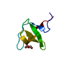
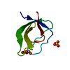
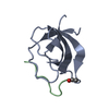
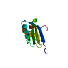
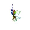
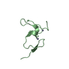
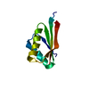
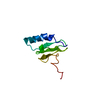
- PDB-4j9f: Crystal structure of the Abl-SH3 domain complexed with the high a... -
+ Open data

- Basic information
| Entry | Database: PDB / ID: 4j9f | ||||||
|---|---|---|---|---|---|---|---|
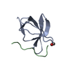
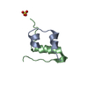
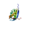
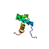
| Title | Crystal structure of the Abl-SH3 domain complexed with the high affinity peptide P0 | ||||||
 Components Components |
| ||||||
 Keywords Keywords | Transferase/unknown function / beta shandwich /  SH3 domain / kinase / poly proline rich motifs / Transferase / Transferase-unknown function complex SH3 domain / kinase / poly proline rich motifs / Transferase / Transferase-unknown function complex | ||||||
| Function / homology |  Function and homology information Function and homology informationregulation of actin filament depolymerization / negative regulation of small GTPase mediated signal transduction / semaphorin receptor binding / negative regulation of phospholipase C activity / positive regulation of actin filament binding / positive regulation of oxidoreductase activity / transitional one stage B cell differentiation / protein localization to cytoplasmic microtubule plus-end / DNA conformation change / podocyte apoptotic process ...regulation of actin filament depolymerization / negative regulation of small GTPase mediated signal transduction / semaphorin receptor binding / negative regulation of phospholipase C activity / positive regulation of actin filament binding / positive regulation of oxidoreductase activity / transitional one stage B cell differentiation / protein localization to cytoplasmic microtubule plus-end / DNA conformation change / podocyte apoptotic process / DN4 thymocyte differentiation / Role of ABL in ROBO-SLIT signaling / response to epinephrine / activation of protein kinase C activity / nicotinate-nucleotide adenylyltransferase activity / ruffle assembly / regulation of modification of synaptic structure / regulation of Rac protein signal transduction / positive regulation of microtubule binding / delta-catenin binding / B cell proliferation involved in immune response / positive regulation of extracellular matrix organization / neuroepithelial cell differentiation / microspike assembly / positive regulation of Wnt signaling pathway, planar cell polarity pathway / cerebellum morphogenesis / positive regulation of blood vessel branching / regulation of blood vessel endothelial cell migration / B-1 B cell homeostasis / mitochondrial depolarization / negative regulation of ubiquitin-protein transferase activity / neuropilin signaling pathway / neuropilin binding / bubble DNA binding / negative regulation of protein serine/threonine kinase activity / cell junction assembly / filopodium assembly / activated T cell proliferation / cellular response to dopamine / establishment of epithelial cell apical/basal polarity / regulation of cell motility / regulation of Cdc42 protein signal transduction / proline-rich region binding / positive regulation of dendrite development / mitogen-activated protein kinase binding / myoblast proliferation / regulation of small GTPase mediated signal transduction / alpha-beta T cell differentiation / regulation of hematopoietic stem cell differentiation / syntaxin binding / cardiac muscle cell proliferation / regulation of T cell differentiation / regulation of axon extension / HDR through Single Strand Annealing (SSA) / phagocytic cup / phagocytosis, engulfment / negative regulation of double-strand break repair via homologous recombination / positive regulation of cell migration involved in sprouting angiogenesis / Fc-gamma receptor signaling pathway involved in phagocytosis / negative regulation of cell-cell adhesion / Myogenesis / regulation of microtubule polymerization / positive regulation of osteoblast proliferation / cell leading edge / RUNX2 regulates osteoblast differentiation / platelet-derived growth factor receptor-beta signaling pathway / positive regulation of focal adhesion assembly / negative regulation of cellular senescence / Bergmann glial cell differentiation / associative learning / semaphorin-plexin signaling pathway / neuromuscular process controlling balance / regulation of endocytosis / actin monomer binding / negative regulation of BMP signaling pathway / negative regulation of mitotic cell cycle / negative regulation of long-term synaptic potentiation / mismatch repair / endothelial cell migration / RHO GTPases Activate WASPs and WAVEs / bicellular tight junction / positive regulation of T cell migration / canonical NF-kappaB signal transduction / BMP signaling pathway / negative regulation of endothelial cell apoptotic process / regulation of cell adhesion / four-way junction DNA binding / signal transduction in response to DNA damage / positive regulation of substrate adhesion-dependent cell spreading / peptidyl-tyrosine autophosphorylation / positive regulation of vasoconstriction / positive regulation of stress fiber assembly / spleen development / ERK1 and ERK2 cascade / ruffle / cellular response to transforming growth factor beta stimulus / RAC1 GTPase cycle / positive regulation of establishment of T cell polarity / actin filament polymerization / positive regulation of interleukin-2 productionSimilarity search - Function | ||||||
| Biological species |  Homo sapiens (human) Homo sapiens (human) | ||||||
| Method | X-RAY DIFFRACTION / SYNCHROTRON / MOLECULAR REPLACEMENT / molecular replacement / Resolution: 1.094 Å | ||||||
 Authors Authors | Camara-Artigas, A. | ||||||
 Citation Citation | Journal: To be Published Title: Crystal structure of the Abl-SH3 domain complexed with the high affinity peptide P0 Authors: Camara-Artigas, A. #1: Journal: Acta Crystallogr.,Sect.D / Year: 2007Title: Crystallization by capillary counter-diffusion and structure determination of the N114A mutant of the SH3 domain of Abl tyrosine kinase complexed with a high-affinity peptide ligand. Authors: Camara-Artigas, A. / Palencia, A. / Martinez, J.C. / Luque, I. / Gavira, J.A. / Garcia-Ruiz, J.M. #2: Journal: J.Biol.Chem. / Year: 2010Title: Role of interfacial water molecules in proline-rich ligand recognition by the Src homology 3 domain of Abl. Authors: Palencia, A. / Camara-Artigas, A. / Pisabarro, M.T. / Martinez, J.C. / Luque, I. | ||||||
| History |
|
- Structure visualization
| Structure viewer | Molecule: MolmilJmol/JSmol |
|---|
- Downloads & links
-Download
| PDBx/mmCIF format | 4j9f.cif.gz | 126.9 KB | Display | PDBx/mmCIF format |
|---|---|---|---|---|
| PDB format | pdb4j9f.ent.gz | 100.6 KB | Display | PDB format |
| PDBx/mmJSON format | 4j9f.json.gz | Tree view | PDBx/mmJSON format | |
| Others |  Other downloads Other downloads |
-Validation report
| Arichive directory | https://data.pdbj.org/pub/pdb/validation_reports/j9/4j9fftp://data.pdbj.org/pub/pdb/validation_reports/j9/4j9f | HTTPS FTP |
|---|
-Related structure data
| Related structure data | 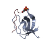 2o88S S: Starting model for refinement |
|---|---|
| Similar structure data |


-Links
 PDBj
PDBj


- Assembly
| Deposited unit | 
| |||||||||||||||
|---|---|---|---|---|---|---|---|---|---|---|---|---|---|---|---|---|
| 1 | 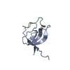
| |||||||||||||||
| 2 | 
| |||||||||||||||
| 3 | 
| |||||||||||||||
| Unit cell |
| |||||||||||||||
| Components on special symmetry positions |
| |||||||||||||||
| Details | biological unit is the complex between one molecule of SH3 domain and the peptide |
-Components
| #1: Protein | Mass: 7009.694 Da / Num. of mol.: 3 / Fragment: SH3 domain (unp residues 60-121) Source method: isolated from a genetically manipulated source Source: (gene. exp.) Homo sapiens (human) / Gene: ABL, ABL1, JTK7 / Plasmid: pET15b / Production host:  Escherichia coli (E. coli) / Strain (production host): BL21(DE3) Escherichia coli (E. coli) / Strain (production host): BL21(DE3)References: UniProt: P00519, non-specific protein-tyrosine kinase#2: Protein/peptide | Mass: 1075.255 Da / Num. of mol.: 3 / Source method: obtained synthetically / References: UniProt: Q9Y3L3*PLUS #3: Chemical | ChemComp-SO4 / | Sulfate  Mass: 96.063 Da / Num. of mol.: 1 / Source method: obtained synthetically / Formula: SO4 Mass: 96.063 Da / Num. of mol.: 1 / Source method: obtained synthetically / Formula: SO4#4: Chemical | ChemComp-GOL / | Glycerol  Mass: 92.094 Da / Num. of mol.: 1 / Source method: obtained synthetically / Formula: C3H8O3 Mass: 92.094 Da / Num. of mol.: 1 / Source method: obtained synthetically / Formula: C3H8O3#5: Water | ChemComp-HOH / | Water Mass: 18.015 Da / Num. of mol.: 222 / Source method: isolated from a natural source / Formula: H2O Mass: 18.015 Da / Num. of mol.: 222 / Source method: isolated from a natural source / Formula: H2O |
|---|
-Experimental details
-Experiment
| Experiment | Method: X-RAY DIFFRACTION / Number of used crystals: 1 |
|---|
- Sample preparation
| Crystal | Density Matthews: 2.01 Å3/Da / Density % sol: 38.73 % |
|---|---|
| Crystal grow | Temperature: 298 K / pH: 7 Details: 2M Ammonium sulphate, 5% PEG300, 0.05M Litium Formate, 10 % glycerol, 0.1M MOPS , pH 7, VAPOR DIFFUSION, SITTING DROP, temperature 298K |
-Data collection
| Diffraction | Mean temperature: 100 K |
|---|---|
| Diffraction source | Source: SYNCHROTRON / Site: ESRF  / Beamline: ID14-4 / Wavelength: 0.97 / Beamline: ID14-4 / Wavelength: 0.97 |
| Detector | Type: ADSC QUANTUM 315r / Detector: CCD / Date: Jan 28, 2011 |
| Radiation | Monochromator: CHANNEL CUT ESRF MONOCHROMATOR AND TORODIAL FOCUSING MIRROR Protocol: SINGLE WAVELENGTH / Monochromatic (M) / Laue (L): M / Scattering type: x-ray |
| Radiation wavelength | Wavelength: 0.97 Å / Relative weight: 1 |
| Reflection | Resolution: 1.094→74.85 Å / Num. obs: 77433 / % possible obs: 97 % / Observed criterion σ(I): 0 / Redundancy: 8.8 % / Biso Wilson estimate: 6.9 Å2 / Rmerge(I) obs: 0.061 / Rsym value: 0.061 / Net I/σ(I): 19.2 |
| Reflection shell | Resolution: 1.09→1.15 Å / Redundancy: 8.7 % / Rmerge(I) obs: 0.43 / Mean I/σ(I) obs: 4.9 / % possible all: 94.9 |
-Phasing
| Phasing | Method: molecular replacement |
|---|
- Processing
| Software |
| |||||||||||||||||||||||||||||||||||||||||||||||||||||||||||||||||||||||||||||||||||||||||||||||||||||||||||||||||||||||||||||||||||||||||||||||||||||||||||||||||||||||||||||||||||||||||||||||||||||||||||
|---|---|---|---|---|---|---|---|---|---|---|---|---|---|---|---|---|---|---|---|---|---|---|---|---|---|---|---|---|---|---|---|---|---|---|---|---|---|---|---|---|---|---|---|---|---|---|---|---|---|---|---|---|---|---|---|---|---|---|---|---|---|---|---|---|---|---|---|---|---|---|---|---|---|---|---|---|---|---|---|---|---|---|---|---|---|---|---|---|---|---|---|---|---|---|---|---|---|---|---|---|---|---|---|---|---|---|---|---|---|---|---|---|---|---|---|---|---|---|---|---|---|---|---|---|---|---|---|---|---|---|---|---|---|---|---|---|---|---|---|---|---|---|---|---|---|---|---|---|---|---|---|---|---|---|---|---|---|---|---|---|---|---|---|---|---|---|---|---|---|---|---|---|---|---|---|---|---|---|---|---|---|---|---|---|---|---|---|---|---|---|---|---|---|---|---|---|---|---|---|---|---|---|---|---|
| Refinement | Method to determine structure: MOLECULAR REPLACEMENT Starting model: 2O88 Resolution: 1.094→21.84 Å / Occupancy max: 1 / Occupancy min: 0.1 / SU ML: 0.09 / σ(F): 0 / Phase error: 13.91 / Stereochemistry target values: ML
| |||||||||||||||||||||||||||||||||||||||||||||||||||||||||||||||||||||||||||||||||||||||||||||||||||||||||||||||||||||||||||||||||||||||||||||||||||||||||||||||||||||||||||||||||||||||||||||||||||||||||||
| Solvent computation | Shrinkage radii: 0.9 Å / VDW probe radii: 1.11 Å / Solvent model: FLAT BULK SOLVENT MODEL | |||||||||||||||||||||||||||||||||||||||||||||||||||||||||||||||||||||||||||||||||||||||||||||||||||||||||||||||||||||||||||||||||||||||||||||||||||||||||||||||||||||||||||||||||||||||||||||||||||||||||||
| Displacement parameters | Biso mean: 13.54 Å2 | |||||||||||||||||||||||||||||||||||||||||||||||||||||||||||||||||||||||||||||||||||||||||||||||||||||||||||||||||||||||||||||||||||||||||||||||||||||||||||||||||||||||||||||||||||||||||||||||||||||||||||
| Refinement step | Cycle: LAST / Resolution: 1.094→21.84 Å
| |||||||||||||||||||||||||||||||||||||||||||||||||||||||||||||||||||||||||||||||||||||||||||||||||||||||||||||||||||||||||||||||||||||||||||||||||||||||||||||||||||||||||||||||||||||||||||||||||||||||||||
| Refine LS restraints |
| |||||||||||||||||||||||||||||||||||||||||||||||||||||||||||||||||||||||||||||||||||||||||||||||||||||||||||||||||||||||||||||||||||||||||||||||||||||||||||||||||||||||||||||||||||||||||||||||||||||||||||
| LS refinement shell |
|
