 Movie
Movie Controller

[English] 日本語
 Yorodumi
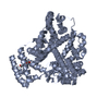
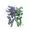
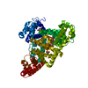
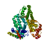
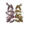
- PDB-4j2v: Crystal Structure of Equine Serum Albumin in complex with 3,5-dii... -
+ Open data

- Basic information
| Entry | Database: PDB / ID: 4j2v | ||||||
|---|---|---|---|---|---|---|---|
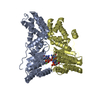
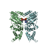
| Title | Crystal Structure of Equine Serum Albumin in complex with 3,5-diiodosalicylic acid | ||||||
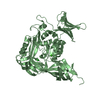
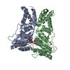
 Components Components | Serum albumin | ||||||
 Keywords Keywords | TRANSPORT PROTEIN / Equine serum albumin / Helical / Serum Albumin Superfamily / Transport / Fatty acids / metabolites and drugs / Plasma | ||||||
| Function / homology |  Function and homology information Function and homology informationcellular response to calcium ion starvation / enterobactin binding / negative regulation of mitochondrial depolarization / toxic substance binding / cellular response to starvation / fatty acid binding / pyridoxal phosphate binding / protein-containing complex / DNA binding / extracellular space ...cellular response to calcium ion starvation / enterobactin binding / negative regulation of mitochondrial depolarization / toxic substance binding / cellular response to starvation / fatty acid binding / pyridoxal phosphate binding / protein-containing complex / DNA binding / extracellular space / metal ion binding / cytoplasmSimilarity search - Function | ||||||
| Biological species |  Equus caballus (horse) Equus caballus (horse) | ||||||
| Method | X-RAY DIFFRACTION / SYNCHROTRON / MOLECULAR REPLACEMENT / Resolution: 2.12 Å | ||||||
 Authors Authors | Sekula, B. / Bujacz, A. / Zielinski, K. / Bujacz, G. | ||||||
 Citation Citation | Journal: Int.J.Biol.Macromol. / Year: 2013 Title: Crystallographic studies of the complexes of bovine and equine serum albumin with 3,5-diiodosalicylic acid. Authors: Sekula, B. / Zielinski, K. / Bujacz, A. | ||||||
| History |
|
- Structure visualization
| Structure viewer | Molecule: MolmilJmol/JSmol |
|---|
- Downloads & links
-Download
| PDBx/mmCIF format | 4j2v.cif.gz | 251.1 KB | Display | PDBx/mmCIF format |
|---|---|---|---|---|
| PDB format | pdb4j2v.ent.gz | 203.5 KB | Display | PDB format |
| PDBx/mmJSON format | 4j2v.json.gz | Tree view | PDBx/mmJSON format | |
| Others |  Other downloads Other downloads |
-Validation report
| Arichive directory | https://data.pdbj.org/pub/pdb/validation_reports/j2/4j2vftp://data.pdbj.org/pub/pdb/validation_reports/j2/4j2v | HTTPS FTP |
|---|
-Related structure data
| Related structure data |  4jk4C  4f5uS S: Starting model for refinement C: citing same article ( |
|---|---|
| Similar structure data |


-Links
 PDBj
PDBj


- Assembly
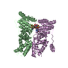
| Deposited unit | 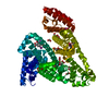
| ||||||||
|---|---|---|---|---|---|---|---|---|---|
| 1 |
| ||||||||
| Unit cell |
|
-Components
-Protein , 1 types, 1 molecules A
| #1: Protein | Mass: 65854.203 Da / Num. of mol.: 1 / Fragment: UNP residues 25-607 / Source method: isolated from a natural source / Details: Plasma / Source: (natural) Equus caballus (horse) / References: UniProt: P35747 |
|---|
-Non-polymers , 5 types, 251 molecules 


| #2: Chemical | ChemComp-DIU /  Mass: 389.914 Da / Num. of mol.: 4 / Source method: obtained synthetically / Formula: C7H4I2O3 Mass: 389.914 Da / Num. of mol.: 4 / Source method: obtained synthetically / Formula: C7H4I2O3#3: Chemical | ChemComp-MLI / Malonic acid Mass: 102.046 Da / Num. of mol.: 7 / Source method: obtained synthetically / Formula: C3H2O4 Mass: 102.046 Da / Num. of mol.: 7 / Source method: obtained synthetically / Formula: C3H2O4#4: Chemical | ChemComp-FMT / Formic acid Mass: 46.025 Da / Num. of mol.: 8 / Source method: obtained synthetically / Formula: CH2O2 Mass: 46.025 Da / Num. of mol.: 8 / Source method: obtained synthetically / Formula: CH2O2#5: Chemical | ChemComp-ACT / | Acetate Mass: 59.044 Da / Num. of mol.: 1 / Source method: obtained synthetically / Formula: C2H3O2 Mass: 59.044 Da / Num. of mol.: 1 / Source method: obtained synthetically / Formula: C2H3O2#6: Water | ChemComp-HOH / | WaterMass: 18.015 Da / Num. of mol.: 231 / Source method: isolated from a natural source / Formula: H2O |
|---|
-Experimental details
-Experiment
| Experiment | Method: X-RAY DIFFRACTION / Number of used crystals: 1 |
|---|
- Sample preparation
| Crystal | Density Matthews: 2.33 Å3/Da / Density % sol: 47.12 % |
|---|---|
| Crystal grow | Temperature: 293 K / Method: vapor diffusion, hanging drop / pH: 6 Details: 85% Tacsimate, cocrystallization with 3,5-diiodosalicylic acid, pH 6.0, VAPOR DIFFUSION, HANGING DROP, temperature 293K |
-Data collection
| Diffraction | Mean temperature: 100 K |
|---|---|
| Diffraction source | Source: SYNCHROTRON / Site: EMBL/DESY, HAMBURG  / Beamline: X12 / Wavelength: 0.932 Å / Beamline: X12 / Wavelength: 0.932 Å |
| Detector | Type: MARMOSAIC 225 mm CCD / Detector: CCD / Date: Aug 21, 2011 / Details: Mirrors |
| Radiation | Monochromator: Double crystal monochromator Si (111) / Protocol: SINGLE WAVELENGTH / Monochromatic (M) / Laue (L): M / Scattering type: x-ray |
| Radiation wavelength | Wavelength: 0.932 Å / Relative weight: 1 |
| Reflection | Resolution: 2.12→33.39 Å / Num. all: 34046 / Num. obs: 34024 / % possible obs: 99.9 % / Observed criterion σ(F): 0 / Observed criterion σ(I): -3 / Redundancy: 5.75 % / Biso Wilson estimate: 43.317 Å2 / Rmerge(I) obs: 0.079 / Net I/σ(I): 16.67 |
| Reflection shell | Resolution: 2.12→2.22 Å / Redundancy: 5.74 % / Rmerge(I) obs: 0.925 / Mean I/σ(I) obs: 2.48 / Num. unique all: 4379 / % possible all: 99.95 |
- Processing
| Software |
| ||||||||||||||||||||||||||||||||||||||||||||||||||||||||||||||||||||||||||||||||||||||||||||||||||||||||||||||||||||||||||||||||||||||||||||||||||||||||||||||||||||||||||||||||||||||||||||||||||||||||||||||||||||||||||||||||||||||||||||||||||||||||||||||||||||||||||||||||||||||||||||||||||||||||||||||||||||||||||||||||||||||||||||||||||||||||||||||
|---|---|---|---|---|---|---|---|---|---|---|---|---|---|---|---|---|---|---|---|---|---|---|---|---|---|---|---|---|---|---|---|---|---|---|---|---|---|---|---|---|---|---|---|---|---|---|---|---|---|---|---|---|---|---|---|---|---|---|---|---|---|---|---|---|---|---|---|---|---|---|---|---|---|---|---|---|---|---|---|---|---|---|---|---|---|---|---|---|---|---|---|---|---|---|---|---|---|---|---|---|---|---|---|---|---|---|---|---|---|---|---|---|---|---|---|---|---|---|---|---|---|---|---|---|---|---|---|---|---|---|---|---|---|---|---|---|---|---|---|---|---|---|---|---|---|---|---|---|---|---|---|---|---|---|---|---|---|---|---|---|---|---|---|---|---|---|---|---|---|---|---|---|---|---|---|---|---|---|---|---|---|---|---|---|---|---|---|---|---|---|---|---|---|---|---|---|---|---|---|---|---|---|---|---|---|---|---|---|---|---|---|---|---|---|---|---|---|---|---|---|---|---|---|---|---|---|---|---|---|---|---|---|---|---|---|---|---|---|---|---|---|---|---|---|---|---|---|---|---|---|---|---|---|---|---|---|---|---|---|---|---|---|---|---|---|---|---|---|---|---|---|---|---|---|---|---|---|---|---|---|---|---|---|---|---|---|---|---|---|---|---|---|---|---|---|---|---|---|---|---|---|---|---|---|---|---|---|---|---|---|---|---|---|---|---|---|---|---|---|---|---|---|---|---|---|---|---|---|---|---|---|---|---|---|---|---|---|---|---|---|---|---|---|---|---|---|---|---|---|---|---|
| Refinement | Method to determine structure: MOLECULAR REPLACEMENT Starting model: 4F5U Resolution: 2.12→33.39 Å / SU ML: 0.3 / Isotropic thermal model: Isotropic / Cross valid method: THROUGHOUT / σ(F): 1.36 / Phase error: 27.14 / Stereochemistry target values: ML
| ||||||||||||||||||||||||||||||||||||||||||||||||||||||||||||||||||||||||||||||||||||||||||||||||||||||||||||||||||||||||||||||||||||||||||||||||||||||||||||||||||||||||||||||||||||||||||||||||||||||||||||||||||||||||||||||||||||||||||||||||||||||||||||||||||||||||||||||||||||||||||||||||||||||||||||||||||||||||||||||||||||||||||||||||||||||||||||||
| Solvent computation | Shrinkage radii: 0.8 Å / VDW probe radii: 1 Å / Solvent model: FLAT BULK SOLVENT MODEL | ||||||||||||||||||||||||||||||||||||||||||||||||||||||||||||||||||||||||||||||||||||||||||||||||||||||||||||||||||||||||||||||||||||||||||||||||||||||||||||||||||||||||||||||||||||||||||||||||||||||||||||||||||||||||||||||||||||||||||||||||||||||||||||||||||||||||||||||||||||||||||||||||||||||||||||||||||||||||||||||||||||||||||||||||||||||||||||||
| Displacement parameters | Biso mean: 37.67 Å2 | ||||||||||||||||||||||||||||||||||||||||||||||||||||||||||||||||||||||||||||||||||||||||||||||||||||||||||||||||||||||||||||||||||||||||||||||||||||||||||||||||||||||||||||||||||||||||||||||||||||||||||||||||||||||||||||||||||||||||||||||||||||||||||||||||||||||||||||||||||||||||||||||||||||||||||||||||||||||||||||||||||||||||||||||||||||||||||||||
| Refinement step | Cycle: LAST / Resolution: 2.12→33.39 Å
| ||||||||||||||||||||||||||||||||||||||||||||||||||||||||||||||||||||||||||||||||||||||||||||||||||||||||||||||||||||||||||||||||||||||||||||||||||||||||||||||||||||||||||||||||||||||||||||||||||||||||||||||||||||||||||||||||||||||||||||||||||||||||||||||||||||||||||||||||||||||||||||||||||||||||||||||||||||||||||||||||||||||||||||||||||||||||||||||
| Refine LS restraints |
| ||||||||||||||||||||||||||||||||||||||||||||||||||||||||||||||||||||||||||||||||||||||||||||||||||||||||||||||||||||||||||||||||||||||||||||||||||||||||||||||||||||||||||||||||||||||||||||||||||||||||||||||||||||||||||||||||||||||||||||||||||||||||||||||||||||||||||||||||||||||||||||||||||||||||||||||||||||||||||||||||||||||||||||||||||||||||||||||
| LS refinement shell |
| ||||||||||||||||||||||||||||||||||||||||||||||||||||||||||||||||||||||||||||||||||||||||||||||||||||||||||||||||||||||||||||||||||||||||||||||||||||||||||||||||||||||||||||||||||||||||||||||||||||||||||||||||||||||||||||||||||||||||||||||||||||||||||||||||||||||||||||||||||||||||||||||||||||||||||||||||||||||||||||||||||||||||||||||||||||||||||||||
| Refinement TLS params. | Method: refined / Refine-ID: X-RAY DIFFRACTION
| ||||||||||||||||||||||||||||||||||||||||||||||||||||||||||||||||||||||||||||||||||||||||||||||||||||||||||||||||||||||||||||||||||||||||||||||||||||||||||||||||||||||||||||||||||||||||||||||||||||||||||||||||||||||||||||||||||||||||||||||||||||||||||||||||||||||||||||||||||||||||||||||||||||||||||||||||||||||||||||||||||||||||||||||||||||||||||||||
| Refinement TLS group |
|
