 Movie
Movie Controller


+ Open data

- Basic information
| Entry | Database: PDB / ID: 4j2l | ||||||
|---|---|---|---|---|---|---|---|
| Title | Crystal Structure of AXH domain complexed with Capicua | ||||||
 Components Components |
| ||||||
 Keywords Keywords |  TRANSCRIPTION REGULATOR / AXH domain / homodimerization protein-protein interaction / Capciua / ATXN1 TRANSCRIPTION REGULATOR / AXH domain / homodimerization protein-protein interaction / Capciua / ATXN1 | ||||||
| Function / homology |  Function and homology information Function and homology informationpoly(G) binding / nuclear inclusion body / nuclear export / poly(U) RNA binding / social behavior / RNA processing / learning / RNA polymerase II transcription regulatory region sequence-specific DNA binding / brain development / memory ...poly(G) binding / nuclear inclusion body / nuclear export / poly(U) RNA binding / social behavior / RNA processing / learning / RNA polymerase II transcription regulatory region sequence-specific DNA binding / brain development / memory / nuclear matrix / : / nervous system development / DNA-binding transcription factor activity, RNA polymerase II-specific / intracellular membrane-bounded organelle / negative regulation of DNA-templated transcription / chromatin / nucleolus / regulation of transcription by RNA polymerase II / negative regulation of transcription by RNA polymerase II / DNA binding / nucleoplasm / identical protein binding / nucleus / cytosol / cytoplasmSimilarity search - Function | ||||||
| Biological species |  Homo sapiens (human) Homo sapiens (human) | ||||||
| Method | X-RAY DIFFRACTION / SYNCHROTRON / MOLECULAR REPLACEMENT / Resolution: 3.15 Å | ||||||
 Authors Authors | Song, J.-J. / Kim, E. | ||||||
 Citation Citation | Journal: Genes Dev. / Year: 2013 Title: Structural basis of protein complex formation and reconfiguration by polyglutamine disease protein Ataxin-1 and Capicua Authors: Kim, E. / Lu, H.-C. / Zoghbi, H.Y. / Song, J.-J. | ||||||
| History |
|
- Structure visualization
| Structure viewer | Molecule: MolmilJmol/JSmol |
|---|
- Downloads & links
-Download
| PDBx/mmCIF format | 4j2l.cif.gz | 64.1 KB | Display | PDBx/mmCIF format |
|---|---|---|---|---|
| PDB format | pdb4j2l.ent.gz | 48.2 KB | Display | PDB format |
| PDBx/mmJSON format | 4j2l.json.gz | Tree view | PDBx/mmJSON format | |
| Others |  Other downloads Other downloads |
-Validation report
| Arichive directory | https://data.pdbj.org/pub/pdb/validation_reports/j2/4j2lftp://data.pdbj.org/pub/pdb/validation_reports/j2/4j2l | HTTPS FTP |
|---|
-Related structure data
| Related structure data |  4j2jSC S: Starting model for refinement C: citing same article ( |
|---|---|
| Similar structure data |


-Links
 PDBj
PDBj

- Assembly
| Deposited unit | 
| ||||||||
|---|---|---|---|---|---|---|---|---|---|
| 1 | 
| ||||||||
| 2 | 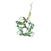
| ||||||||
| Unit cell |
|
-Components
| #1: Protein | Ataxin 1 / Spinocerebellar ataxia type 1 protein Mass: 14119.982 Da / Num. of mol.: 2 / Fragment: AXH domain, UNP residues 562-688 Source method: isolated from a genetically manipulated source Source: (gene. exp.) Homo sapiens (human) / Gene: ATXN1 / Production host:  Escherichia coli (E. coli) / References: UniProt: P54253 Escherichia coli (E. coli) / References: UniProt: P54253#2: Protein/peptide | Mass: 3244.760 Da / Num. of mol.: 2 / Fragment: UNP residues 21-48 Source method: isolated from a genetically manipulated source Source: (gene. exp.) Homo sapiens (human) / Gene: CIC / Production host: Escherichia coli (E. coli) / References: UniProt: Q96RK0 |
|---|
-Experimental details
-Experiment
| Experiment | Method: X-RAY DIFFRACTION / Number of used crystals: 1 |
|---|
- Sample preparation
| Crystal | Density Matthews: 2.85 Å3/Da / Density % sol: 56.77 % |
|---|---|
| Crystal grow | Temperature: 293 K / Method: vapor diffusion, hanging drop / pH: 8 Details: 1.6M NaCl, 3%(v/v) glycerol, 16%(w/v) PEG3350, 1mM L-Glutathione, pH 8.0, VAPOR DIFFUSION, HANGING DROP, temperature 293K |
-Data collection
| Diffraction | Mean temperature: 100 K |
|---|---|
| Diffraction source | Source: SYNCHROTRON / Site: PAL/PLS  / Beamline: 6B / Beamline: 6B |
| Detector | Type: ADSC QUANTUM 315r / Detector: CCD / Date: Jul 19, 2012 |
| Radiation | Protocol: SINGLE WAVELENGTH / Monochromatic (M) / Laue (L): M / Scattering type: x-ray |
| Radiation wavelength | Relative weight: 1 |
| Reflection | Resolution: 3→50 Å / Num. obs: 8720 / % possible obs: 99.9 % |
| Reflection shell | Resolution: 3→3.05 Å / Redundancy: 13.3 % / Rmerge(I) obs: 0.663 / Mean I/σ(I) obs: 6.2 / % possible all: 100 |
- Processing
| Software |
| |||||||||||||||||||||||||||
|---|---|---|---|---|---|---|---|---|---|---|---|---|---|---|---|---|---|---|---|---|---|---|---|---|---|---|---|---|
| Refinement | Method to determine structure: MOLECULAR REPLACEMENT Starting model: 4J2J Resolution: 3.15→29.23 Å / Rfactor Rfree error: 0.014 / Data cutoff high absF: 107840.74 / Data cutoff low absF: 0 / Isotropic thermal model: RESTRAINED / Cross valid method: THROUGHOUT / σ(F): 0 / Details: BULK SOLVENT MODEL USED
| |||||||||||||||||||||||||||
| Solvent computation | Solvent model: FLAT MODEL / Bsol: 53.9275 Å2 / ksol: 0.35 e/Å3 | |||||||||||||||||||||||||||
| Displacement parameters | Biso mean: 75.2161 Å2
| |||||||||||||||||||||||||||
| Refine analyze |
| |||||||||||||||||||||||||||
| Refinement step | Cycle: LAST / Resolution: 3.15→29.23 Å
| |||||||||||||||||||||||||||
| Refine LS restraints |
| |||||||||||||||||||||||||||
| LS refinement shell | Resolution: 3.15→3.35 Å / Rfactor Rfree error: 0.041 / Total num. of bins used: 6
| |||||||||||||||||||||||||||
| Xplor file | Serial no: 1 / Param file: protein_rep.param / Topol file: protein.top |
