 Movie
Movie Controller

[English] 日本語
 Yorodumi
- PDB-2edo: Solution structure of the first ig-like domain from human CD48 antigen -
+ Open data

- Basic information
| Entry | Database: PDB / ID: 2edo | ||||||
|---|---|---|---|---|---|---|---|
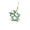
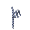
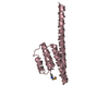
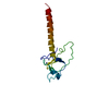
| Title | Solution structure of the first ig-like domain from human CD48 antigen | ||||||
 Components Components | CD48 antigen CD48 CD48 | ||||||
 Keywords Keywords | CELL ADHESION / beta-sandwich / ig-fold / B-lymphocyte activation marker BLAST-1 / BCM1 surface antigen / Leukocyte antigen MEM-102 / TCT.1 / Structural Genomics / NPPSFA / National Project on Protein Structural and Functional Analyses / RIKEN Structural Genomics/Proteomics Initiative / RSGI | ||||||
| Function / homology |  Function and homology information: / regulation of adaptive immune response / : / leukocyte migration / antigen binding / Cell surface interactions at the vascular wall / defense response / signaling receptor activity / membrane raft / extracellular exosome ...: / regulation of adaptive immune response / : / leukocyte migration / antigen binding / Cell surface interactions at the vascular wall / defense response / signaling receptor activity / membrane raft / extracellular exosome / membrane / plasma membrane Function and homology information: / regulation of adaptive immune response / : / leukocyte migration / antigen binding / Cell surface interactions at the vascular wall / defense response / signaling receptor activity / membrane raft / extracellular exosome ...: / regulation of adaptive immune response / : / leukocyte migration / antigen binding / Cell surface interactions at the vascular wall / defense response / signaling receptor activity / membrane raft / extracellular exosome / membrane / plasma membraneSimilarity search - Function | ||||||
| Biological species |  Homo sapiens (human) Homo sapiens (human) | ||||||
| Method | SOLUTION NMR / torsion angle dynamics | ||||||
 Authors Authors | Nagashima, K. / Nagashima, T. / Yoshida, M. / Hayashi, F. / Yokoyama, S. / RIKEN Structural Genomics/Proteomics Initiative (RSGI) | ||||||
 Citation Citation | Journal: To be Published Title: Solution structure of the first ig-like domain from human CD48 antigen Authors: Nagashima, K. / Nagashima, T. / Yoshida, M. / Hayashi, F. / Yokoyama, S. | ||||||
| History |
|
- Structure visualization
| Structure viewer | Molecule: MolmilJmol/JSmol |
|---|
- Downloads & links
-Download
| PDBx/mmCIF format | 2edo.cif.gz | 768.7 KB | Display | PDBx/mmCIF format |
|---|---|---|---|---|
| PDB format | pdb2edo.ent.gz | 646.2 KB | Display | PDB format |
| PDBx/mmJSON format | 2edo.json.gz | Tree view | PDBx/mmJSON format | |
| Others |  Other downloads Other downloads |
-Validation report
| Arichive directory | https://data.pdbj.org/pub/pdb/validation_reports/ed/2edoftp://data.pdbj.org/pub/pdb/validation_reports/ed/2edo | HTTPS FTP |
|---|
-Related structure data
| Similar structure data | |
|---|---|
| Other databases |


-Links
 PDBj
PDBj
- Assembly
| Deposited unit | 
| |||||||||
|---|---|---|---|---|---|---|---|---|---|---|
| 1 |
| |||||||||
| NMR ensembles |
|
-Components
| #1: Protein | CD48 / B-lymphocyte activation marker BLAST-1 / BCM1 surface antigen / Leukocyte antigen MEM-102 / TCT.1 Mass: 13901.872 Da / Num. of mol.: 1 / Fragment: V-set Source method: isolated from a genetically manipulated source Source: (gene. exp.) Homo sapiens (human) / Description: cell free protein synthesis / Gene: CD48, BCM1, BLAST1 / Plasmid: P060411-11 / References: UniProt: P09326 |
|---|
-Experimental details
-Experiment
| Experiment | Method: SOLUTION NMR | ||||||||||||
|---|---|---|---|---|---|---|---|---|---|---|---|---|---|
| NMR experiment |
| ||||||||||||
| NMR details | Text: spectrometer_id 1 for 3D_13C-separated_NOESY; spectrometer_id 2 for 3D_15N-separated_NOESY |
- Sample preparation
| Details | Contents: 1.15mM 13C, 15N-labeled protein; 20mM d-Tris-HCl(pH7.0); 100mM NaCl 1mM d-DTT; 0.02% NaN3, 10% D2O; 90% H2O Solvent system: 10% D2O; 90% H2O |
|---|---|
| Sample conditions | Ionic strength: 120mM / pH: 7.0 / Pressure: ambient / Temperature: 293 K |
-NMR measurement
| NMR spectrometer |
|
|---|
- Processing
| NMR software |
| ||||||||||||||||||||||||||||
|---|---|---|---|---|---|---|---|---|---|---|---|---|---|---|---|---|---|---|---|---|---|---|---|---|---|---|---|---|---|
| Refinement | Method: torsion angle dynamics / Software ordinal: 1 | ||||||||||||||||||||||||||||
| NMR representative | Selection criteria: lowest energy | ||||||||||||||||||||||||||||
| NMR ensemble | Conformer selection criteria: structures with the least restraint violations, target function Conformers calculated total number: 100 / Conformers submitted total number: 20 |
