 Movie
Movie Controller


+ Open data

- Basic information
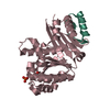
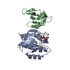
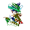
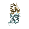
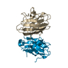
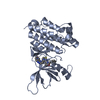
| Entry | Database: PDB / ID: 4j2g | ||||||
|---|---|---|---|---|---|---|---|
| Title | Atg13 HORMA domain | ||||||
 Components Components | KLTH0A00704p | ||||||
 Keywords Keywords | UNKNOWN FUNCTION /  Autophagy / HORMA / Atg13 Autophagy / HORMA / Atg13 | ||||||
| Function / homology |  Function and homology information Function and homology informationAtg1/ULK1 kinase complex / protein localization to phagophore assembly site / piecemeal microautophagy of the nucleus / phagophore assembly site / mitophagy / cytosolSimilarity search - Function | ||||||
| Biological species |  Lachancea thermotolerans (fungus) Lachancea thermotolerans (fungus) | ||||||
| Method | X-RAY DIFFRACTION / SYNCHROTRON / SAD / Resolution: 2.29 Å | ||||||
 Authors Authors | Jao, C. / Stanley, R.E. / Ragusa, M.J. / Hurley, J.H. | ||||||
 Citation Citation | Journal: Proc.Natl.Acad.Sci.USA / Year: 2013 Title: A HORMA domain in Atg13 mediates PI 3-kinase recruitment in autophagy. Authors: Jao, C.C. / Ragusa, M.J. / Stanley, R.E. / Hurley, J.H. | ||||||
| History |
|
- Structure visualization
| Structure viewer | Molecule: MolmilJmol/JSmol |
|---|
- Downloads & links
-Download
| PDBx/mmCIF format | 4j2g.cif.gz | 89.9 KB | Display | PDBx/mmCIF format |
|---|---|---|---|---|
| PDB format | pdb4j2g.ent.gz | 73.4 KB | Display | PDB format |
| PDBx/mmJSON format | 4j2g.json.gz | Tree view | PDBx/mmJSON format | |
| Others |  Other downloads Other downloads |
-Validation report
| Arichive directory | https://data.pdbj.org/pub/pdb/validation_reports/j2/4j2gftp://data.pdbj.org/pub/pdb/validation_reports/j2/4j2g | HTTPS FTP |
|---|
-Related structure data
| Similar structure data |
|---|


-Links
 PDBj
PDBj
- Assembly
| Deposited unit | 
| ||||||||
|---|---|---|---|---|---|---|---|---|---|
| 1 | 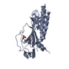
| ||||||||
| 2 | 
| ||||||||
| 3 |
| ||||||||
| Unit cell |
|
-Components
| #1: Protein | Mass: 30507.037 Da / Num. of mol.: 2 / Fragment: UNP residues 1-260 / Mutation: L123M Source method: isolated from a genetically manipulated source Source: (gene. exp.) Lachancea thermotolerans (fungus) / Strain: ATCC 56472 / CBS 6340 / NRRL Y-8284 / Gene: KLTH0A00704g / Production host:  Escherichia coli (E. coli) / References: UniProt: C5DB94 Escherichia coli (E. coli) / References: UniProt: C5DB94#2: Chemical | Sulfate  Mass: 96.063 Da / Num. of mol.: 2 / Source method: obtained synthetically / Formula: SO4 Mass: 96.063 Da / Num. of mol.: 2 / Source method: obtained synthetically / Formula: SO4#3: Water | ChemComp-HOH / | Water Mass: 18.015 Da / Num. of mol.: 91 / Source method: isolated from a natural source / Formula: H2O Mass: 18.015 Da / Num. of mol.: 91 / Source method: isolated from a natural source / Formula: H2O |
|---|
-Experimental details
-Experiment
| Experiment | Method: X-RAY DIFFRACTION / Number of used crystals: 1 |
|---|
- Sample preparation
| Crystal | Density Matthews: 2.68 Å3/Da / Density % sol: 54.16 % |
|---|---|
| Crystal grow | Temperature: 294 K / Method: vapor diffusion, hanging drop / pH: 6.5 Details: 0.2mM ammonium sulfate, 0.1M MES pH6.5, 20% w/v PEG 8000, VAPOR DIFFUSION, HANGING DROP, temperature 294K |
-Data collection
| Diffraction | Mean temperature: 200 K |
|---|---|
| Diffraction source | Source: SYNCHROTRON / Site: Diamond  / Beamline: I02 / Wavelength: 1 Å / Beamline: I02 / Wavelength: 1 Å |
| Detector | Type: PSI PILATUS 6M / Detector: PIXEL / Date: May 28, 2012 |
| Radiation | Monochromator: filter / Protocol: SINGLE WAVELENGTH / Monochromatic (M) / Laue (L): M / Scattering type: x-ray |
| Radiation wavelength | Wavelength: 1 Å / Relative weight: 1 |
| Reflection | Resolution: 2.29→84.05 Å / Num. all: 29497 / % possible obs: 99 % / Observed criterion σ(F): 2.6 / Observed criterion σ(I): 12.1 |
- Processing
| Software |
| ||||||||||||||||||||||||||||||||||||||||||||||||||||||||||||||||||||||||||||||||||||||||||||||||||||||||||||||||||||||||||||||||||||||||||||||||||||||||||||||||||||||||||
|---|---|---|---|---|---|---|---|---|---|---|---|---|---|---|---|---|---|---|---|---|---|---|---|---|---|---|---|---|---|---|---|---|---|---|---|---|---|---|---|---|---|---|---|---|---|---|---|---|---|---|---|---|---|---|---|---|---|---|---|---|---|---|---|---|---|---|---|---|---|---|---|---|---|---|---|---|---|---|---|---|---|---|---|---|---|---|---|---|---|---|---|---|---|---|---|---|---|---|---|---|---|---|---|---|---|---|---|---|---|---|---|---|---|---|---|---|---|---|---|---|---|---|---|---|---|---|---|---|---|---|---|---|---|---|---|---|---|---|---|---|---|---|---|---|---|---|---|---|---|---|---|---|---|---|---|---|---|---|---|---|---|---|---|---|---|---|---|---|---|---|---|
| Refinement | Method to determine structure: SAD / Resolution: 2.29→57.25 Å / Cor.coef. Fo:Fc: 0.951 / Cor.coef. Fo:Fc free: 0.929 / Occupancy max: 1 / Occupancy min: 0.59 / SU B: 6.927 / SU ML: 0.17 / Cross valid method: THROUGHOUT / ESU R: 0.234 / ESU R Free: 0.21 Stereochemistry target values: MAXIMUM LIKELIHOOD WITH PHASES Details: HYDROGENS HAVE BEEN USED IF PRESENT IN THE INPUT
| ||||||||||||||||||||||||||||||||||||||||||||||||||||||||||||||||||||||||||||||||||||||||||||||||||||||||||||||||||||||||||||||||||||||||||||||||||||||||||||||||||||||||||
| Solvent computation | Ion probe radii: 0.8 Å / Shrinkage radii: 0.8 Å / VDW probe radii: 1.2 Å / Solvent model: MASK | ||||||||||||||||||||||||||||||||||||||||||||||||||||||||||||||||||||||||||||||||||||||||||||||||||||||||||||||||||||||||||||||||||||||||||||||||||||||||||||||||||||||||||
| Displacement parameters | Biso mean: 67.413 Å2
| ||||||||||||||||||||||||||||||||||||||||||||||||||||||||||||||||||||||||||||||||||||||||||||||||||||||||||||||||||||||||||||||||||||||||||||||||||||||||||||||||||||||||||
| Refinement step | Cycle: LAST / Resolution: 2.29→57.25 Å
| ||||||||||||||||||||||||||||||||||||||||||||||||||||||||||||||||||||||||||||||||||||||||||||||||||||||||||||||||||||||||||||||||||||||||||||||||||||||||||||||||||||||||||
| Refine LS restraints |
| ||||||||||||||||||||||||||||||||||||||||||||||||||||||||||||||||||||||||||||||||||||||||||||||||||||||||||||||||||||||||||||||||||||||||||||||||||||||||||||||||||||||||||
| LS refinement shell | Resolution: 2.29→2.349 Å / Total num. of bins used: 20
|
