 Movie
Movie Controller

[English] 日本語
 Yorodumi
- PDB-4isl: Crystal Structure of the inactive Matriptase in complex with its ... -
+ Open data

- Basic information
| Entry | Database: PDB / ID: 4isl | ||||||
|---|---|---|---|---|---|---|---|
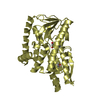
| Title | Crystal Structure of the inactive Matriptase in complex with its inhibitor HAI-1 | ||||||
 Components Components |
| ||||||
 Keywords Keywords | hydrolase/hydrolase inhibitor /  Beta barrel / Serine protease inhibitor / epithelium / hydrolase-hydrolase inhibitor complex Beta barrel / Serine protease inhibitor / epithelium / hydrolase-hydrolase inhibitor complex | ||||||
| Function / homology |  Function and homology information Function and homology informationepithelium development / Signaling by MST1 / matriptase / positive regulation of glial cell differentiation / epithelial cell morphogenesis involved in placental branching / acrosome reaction / negative regulation of neural precursor cell proliferation / branching involved in labyrinthine layer morphogenesis / placenta blood vessel development / Formation of the cornified envelope ...epithelium development / Signaling by MST1 / matriptase / positive regulation of glial cell differentiation / epithelial cell morphogenesis involved in placental branching / acrosome reaction / negative regulation of neural precursor cell proliferation / branching involved in labyrinthine layer morphogenesis / placenta blood vessel development / Formation of the cornified envelope / MET Receptor Activation / cellular response to BMP stimulus / epidermis development / keratinocyte differentiation / serine-type peptidase activity / extracellular matrix organization / neural tube closure / protein catabolic process / serine-type endopeptidase inhibitor activity / basolateral plasma membrane / external side of plasma membrane / serine-type endopeptidase activity / proteolysis / extracellular space / extracellular exosome / extracellular region / membrane / plasma membrane / cytoplasmSimilarity search - Function | ||||||
| Biological species |  Homo sapiens (human) Homo sapiens (human) | ||||||
| Method | X-RAY DIFFRACTION / SYNCHROTRON / MOLECULAR REPLACEMENT / Resolution: 2.29 Å | ||||||
 Authors Authors | Huang, M.D. / Zhao, B.Y. / Yuan, C. / Li, R. | ||||||
 Citation Citation | Journal: J.Biol.Chem. / Year: 2013 Title: Crystal structures of matriptase in complex with its inhibitor hepatocyte growth factor activator inhibitor-1. Authors: Zhao, B. / Yuan, C. / Li, R. / Qu, D. / Huang, M. / Ngo, J.C. | ||||||
| History |
|
- Structure visualization
| Structure viewer | Molecule: MolmilJmol/JSmol |
|---|
- Downloads & links
-Download
| PDBx/mmCIF format | 4isl.cif.gz | 80.2 KB | Display | PDBx/mmCIF format |
|---|---|---|---|---|
| PDB format | pdb4isl.ent.gz | 57.9 KB | Display | PDB format |
| PDBx/mmJSON format | 4isl.json.gz | Tree view | PDBx/mmJSON format | |
| Others |  Other downloads Other downloads |
-Validation report
| Arichive directory | https://data.pdbj.org/pub/pdb/validation_reports/is/4islftp://data.pdbj.org/pub/pdb/validation_reports/is/4isl | HTTPS FTP |
|---|
-Related structure data
| Related structure data |  4is5C  4isnC 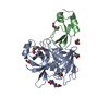 4isoC 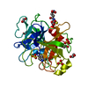 3p8gS C: citing same article ( S: Starting model for refinement |
|---|---|
| Similar structure data |


-Links
 PDBj
PDBj


- Assembly
| Deposited unit | 
| ||||||||
|---|---|---|---|---|---|---|---|---|---|
| 1 |
| ||||||||
| Unit cell |
| ||||||||
| Components on special symmetry positions |
|
-Components
-Protein , 2 types, 2 molecules BA
| #1: Protein | Kunitz domain / Hepatocyte growth factor activator inhibitor type 1 / HAI-1 Mass: 6941.824 Da / Num. of mol.: 1 / Fragment: Kunitz domain I (unp residues 245-304) Source method: isolated from a genetically manipulated source Source: (gene. exp.) Homo sapiens (human) / Gene: HAI1, SPINT1, UNQ223/PRO256 / Plasmid: PMT/BIP/V5-his-A / Production host:  Drosophila melanogaster (fruit fly) / Strain (production host): S2 / References: UniProt: O43278 Drosophila melanogaster (fruit fly) / Strain (production host): S2 / References: UniProt: O43278 |
|---|---|
| #2: Protein | Mass: 26461.783 Da / Num. of mol.: 1 / Fragment: Serine protease domain (unp residues 615-855) / Mutation: N164Q, S805A Source method: isolated from a genetically manipulated source Source: (gene. exp.) Homo sapiens (human) / Gene: PRSS14, SNC19, ST14, TADG15 / Plasmid: pPICZalphaA / Production host:  Komagataella pastoris (fungus) / Strain (production host): X-33 / References: UniProt: Q9Y5Y6, matriptase Komagataella pastoris (fungus) / Strain (production host): X-33 / References: UniProt: Q9Y5Y6, matriptase |
-Non-polymers , 5 types, 178 molecules 


| #3: Chemical | ChemComp-PG4 / Polyethylene glycol Mass: 194.226 Da / Num. of mol.: 1 / Source method: obtained synthetically / Formula: C8H18O5 / Comment: precipitant*YM Mass: 194.226 Da / Num. of mol.: 1 / Source method: obtained synthetically / Formula: C8H18O5 / Comment: precipitant*YM | ||||||
|---|---|---|---|---|---|---|---|
| #4: Chemical | Glycerol Mass: 92.094 Da / Num. of mol.: 3 / Source method: obtained synthetically / Formula: C3H8O3 Mass: 92.094 Da / Num. of mol.: 3 / Source method: obtained synthetically / Formula: C3H8O3#5: Chemical | ChemComp-PGE / | Polyethylene glycol Mass: 150.173 Da / Num. of mol.: 1 / Source method: obtained synthetically / Formula: C6H14O4 Mass: 150.173 Da / Num. of mol.: 1 / Source method: obtained synthetically / Formula: C6H14O4#6: Chemical | ChemComp-GSH / | Glutathione Mass: 307.323 Da / Num. of mol.: 1 / Source method: obtained synthetically / Formula: C10H17N3O6S Mass: 307.323 Da / Num. of mol.: 1 / Source method: obtained synthetically / Formula: C10H17N3O6S#7: Water | ChemComp-HOH / | WaterMass: 18.015 Da / Num. of mol.: 172 / Source method: isolated from a natural source / Formula: H2O |
-Experimental details
-Experiment
| Experiment | Method: X-RAY DIFFRACTION / Number of used crystals: 1 |
|---|
- Sample preparation
| Crystal | Density Matthews: 2.57 Å3/Da / Density % sol: 52.13 % |
|---|---|
| Crystal grow | Temperature: 295 K / Method: vapor diffusion, sitting drop / pH: 8.5 Details: 0.1 M Tris-HCl, 20% (w/v) polyethylene glycol 8000, pH 8.5, VAPOR DIFFUSION, SITTING DROP, temperature 295K |
-Data collection
| Diffraction | Mean temperature: 100 K | |||||||||||||||||||||||||||||||||||||||||||||||||||||||||||||||||||||||||||||||||||||||||||||||||||||||||||||||||||||||||||||||||||||||||||||||||||
|---|---|---|---|---|---|---|---|---|---|---|---|---|---|---|---|---|---|---|---|---|---|---|---|---|---|---|---|---|---|---|---|---|---|---|---|---|---|---|---|---|---|---|---|---|---|---|---|---|---|---|---|---|---|---|---|---|---|---|---|---|---|---|---|---|---|---|---|---|---|---|---|---|---|---|---|---|---|---|---|---|---|---|---|---|---|---|---|---|---|---|---|---|---|---|---|---|---|---|---|---|---|---|---|---|---|---|---|---|---|---|---|---|---|---|---|---|---|---|---|---|---|---|---|---|---|---|---|---|---|---|---|---|---|---|---|---|---|---|---|---|---|---|---|---|---|---|---|---|
| Diffraction source | Source: SYNCHROTRON / Site: SSRF  / Beamline: BL17U / Wavelength: 0.979 Å / Beamline: BL17U / Wavelength: 0.979 Å | |||||||||||||||||||||||||||||||||||||||||||||||||||||||||||||||||||||||||||||||||||||||||||||||||||||||||||||||||||||||||||||||||||||||||||||||||||
| Detector | Type: ADSC QUANTUM 315r / Detector: CCD / Date: Jan 1, 2012 | |||||||||||||||||||||||||||||||||||||||||||||||||||||||||||||||||||||||||||||||||||||||||||||||||||||||||||||||||||||||||||||||||||||||||||||||||||
| Radiation | Protocol: SINGLE WAVELENGTH / Monochromatic (M) / Laue (L): M / Scattering type: x-ray | |||||||||||||||||||||||||||||||||||||||||||||||||||||||||||||||||||||||||||||||||||||||||||||||||||||||||||||||||||||||||||||||||||||||||||||||||||
| Radiation wavelength | Wavelength: 0.979 Å / Relative weight: 1 | |||||||||||||||||||||||||||||||||||||||||||||||||||||||||||||||||||||||||||||||||||||||||||||||||||||||||||||||||||||||||||||||||||||||||||||||||||
| Reflection | Resolution: 2.29→50 Å / Num. obs: 29858 / % possible obs: 98.3 % / Redundancy: 8 % / Rmerge(I) obs: 0.057 / Χ2: 2.247 / Net I/σ(I): 21.1 | |||||||||||||||||||||||||||||||||||||||||||||||||||||||||||||||||||||||||||||||||||||||||||||||||||||||||||||||||||||||||||||||||||||||||||||||||||
| Reflection shell |
|
- Processing
| Software |
| |||||||||||||||||||||||||||||||||||||||||||||||||||||||||||||||||
|---|---|---|---|---|---|---|---|---|---|---|---|---|---|---|---|---|---|---|---|---|---|---|---|---|---|---|---|---|---|---|---|---|---|---|---|---|---|---|---|---|---|---|---|---|---|---|---|---|---|---|---|---|---|---|---|---|---|---|---|---|---|---|---|---|---|---|
| Refinement | Method to determine structure: MOLECULAR REPLACEMENT Starting model: 3P8G Resolution: 2.29→50 Å / Cor.coef. Fo:Fc: 0.942 / Cor.coef. Fo:Fc free: 0.907 / Occupancy max: 1 / Occupancy min: 0.5 / SU B: 5.534 / SU ML: 0.138 / Cross valid method: THROUGHOUT / σ(F): 0 / ESU R: 0.314 / ESU R Free: 0.224 / Stereochemistry target values: MAXIMUM LIKELIHOOD Details: HYDROGENS HAVE BEEN ADDED IN THE RIDING POSITIONS U VALUES : REFINED INDIVIDUALLY
| |||||||||||||||||||||||||||||||||||||||||||||||||||||||||||||||||
| Solvent computation | Ion probe radii: 0.8 Å / Shrinkage radii: 0.8 Å / VDW probe radii: 1.4 Å / Solvent model: MASK | |||||||||||||||||||||||||||||||||||||||||||||||||||||||||||||||||
| Displacement parameters | Biso max: 55.75 Å2 / Biso mean: 21.6666 Å2 / Biso min: 10.3 Å2
| |||||||||||||||||||||||||||||||||||||||||||||||||||||||||||||||||
| Refinement step | Cycle: LAST / Resolution: 2.29→50 Å
| |||||||||||||||||||||||||||||||||||||||||||||||||||||||||||||||||
| Refine LS restraints |
| |||||||||||||||||||||||||||||||||||||||||||||||||||||||||||||||||
| LS refinement shell | Resolution: 2.29→2.35 Å / Total num. of bins used: 20
|
