 Movie
Movie Controller

[English] 日本語
 Yorodumi
- PDB-4ib7: Bovine beta-lactoglobulin (isoform A) in complex with dodecyltrim... -
+ Open data

- Basic information
| Entry | Database: PDB / ID: 4ib7 | ||||||
|---|---|---|---|---|---|---|---|
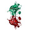
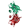
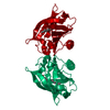
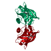
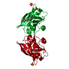
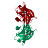
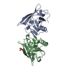
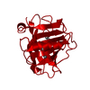
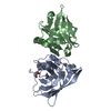
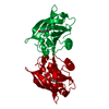
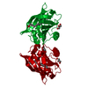
| Title | Bovine beta-lactoglobulin (isoform A) in complex with dodecyltrimethylammonium (DTAC) | ||||||
 Components Components | beta-lactoglobulin | ||||||
 Keywords Keywords | TRANSPORT PROTEIN / lipocalin | ||||||
| Function / homology |  Function and homology informationretinol binding / long-chain fatty acid binding / extracellular space / identical protein binding Function and homology informationretinol binding / long-chain fatty acid binding / extracellular space / identical protein bindingSimilarity search - Function | ||||||
| Biological species |  Bos taurus (cattle) Bos taurus (cattle) | ||||||
| Method | X-RAY DIFFRACTION / MOLECULAR REPLACEMENT / Resolution: 2.2 Å | ||||||
 Authors Authors | Loch, J.I. / Bonarek, P. / Polit, A. / Swiatek, S. / Dziedzicka-Wasylewska, M. / Lewinski, K. | ||||||
 Citation Citation | Journal: J.Mol.Recognit. / Year: 2013 Title: The differences in binding 12-carbon aliphatic ligands by bovine beta-lactoglobulin isoform A and B studied by isothermal titration calorimetry and X-ray crystallography Authors: Loch, J.I. / Bonarek, P. / Polit, A. / Swiatek, S. / Dziedzicka-Wasylewska, M. / Lewinski, K. | ||||||
| History |
| ||||||
| Remark 650 | HELIX DETERMINATION METHOD: AUTHOR DETERMINED |
- Structure visualization
| Structure viewer | Molecule: MolmilJmol/JSmol |
|---|
- Downloads & links
-Download
| PDBx/mmCIF format | 4ib7.cif.gz | 46.2 KB | Display | PDBx/mmCIF format |
|---|---|---|---|---|
| PDB format | pdb4ib7.ent.gz | 31.9 KB | Display | PDB format |
| PDBx/mmJSON format | 4ib7.json.gz | Tree view | PDBx/mmJSON format | |
| Others |  Other downloads Other downloads |
-Validation report
| Arichive directory | https://data.pdbj.org/pub/pdb/validation_reports/ib/4ib7ftp://data.pdbj.org/pub/pdb/validation_reports/ib/4ib7 | HTTPS FTP |
|---|
-Related structure data
| Related structure data |  4ib6C  4ib8C  4ib9C  4ibaC 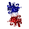 1bsyS C: citing same article ( S: Starting model for refinement |
|---|---|
| Similar structure data |


-Links
 PDBj
PDBj

- Assembly
| Deposited unit | 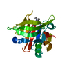
| ||||||||
|---|---|---|---|---|---|---|---|---|---|
| 1 | 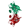
| ||||||||
| Unit cell |
|
-Components
| #1: Protein | / Beta-LG Mass: 18387.264 Da / Num. of mol.: 1 / Fragment: UNP residues 17-178 / Source method: isolated from a natural source / Source: (natural) Bos taurus (cattle) / References: UniProt: P02754 |
|---|---|
| #2: Chemical | ChemComp-CAT /   Mass: 228.437 Da / Num. of mol.: 1 / Source method: obtained synthetically / Formula: C15H34N Mass: 228.437 Da / Num. of mol.: 1 / Source method: obtained synthetically / Formula: C15H34N |
| #3: Water | ChemComp-HOH / Water Mass: 18.015 Da / Num. of mol.: 31 / Source method: isolated from a natural source / Formula: H2O Mass: 18.015 Da / Num. of mol.: 31 / Source method: isolated from a natural source / Formula: H2O |
-Experimental details
-Experiment
| Experiment | Method: X-RAY DIFFRACTION / Number of used crystals: 1 |
|---|
- Sample preparation
| Crystal | Density Matthews: 2.48 Å3/Da / Density % sol: 50.44 % |
|---|---|
| Crystal grow | Temperature: 293 K / Method: vapor diffusion, hanging drop / pH: 7.5 Details: 1.34M TRISODIUM CITRATE, 0.1M TRIS-HCL BUFFER, PH 7.5, VAPOR DIFFUSION, HANGING DROP, temperature 293K |
-Data collection
| Diffraction | Mean temperature: 110 K |
|---|---|
| Diffraction source | Source: SEALED TUBE / Type: OTHER / Wavelength: 1.54 Å |
| Detector | Type: AGILENT ATLAS CCD / Detector: CCD / Date: Jan 25, 2012 |
| Radiation | Protocol: SINGLE WAVELENGTH / Monochromatic (M) / Laue (L): M / Scattering type: x-ray |
| Radiation wavelength | Wavelength: 1.54 Å / Relative weight: 1 |
| Reflection | Resolution: 2.2→14.26 Å / Num. all: 9752 / Num. obs: 9674 / % possible obs: 99.2 % / Observed criterion σ(F): 2 / Observed criterion σ(I): 2 / Rmerge(I) obs: 0.047 / Net I/σ(I): 11.8 |
| Reflection shell | Resolution: 2.2→2.32 Å / Rmerge(I) obs: 0.182 / Mean I/σ(I) obs: 3.3 / Num. unique all: 1420 / % possible all: 100 |
- Processing
| Software |
| |||||||||||||||||||||||||||||||||||||||||||||
|---|---|---|---|---|---|---|---|---|---|---|---|---|---|---|---|---|---|---|---|---|---|---|---|---|---|---|---|---|---|---|---|---|---|---|---|---|---|---|---|---|---|---|---|---|---|---|
| Refinement | Method to determine structure: MOLECULAR REPLACEMENT Starting model: 1BSY Resolution: 2.2→14.26 Å / Cor.coef. Fo:Fc: 0.931 / Cor.coef. Fo:Fc free: 0.881 / SU B: 8.594 / SU ML: 0.211 / Cross valid method: THROUGHOUT / ESU R: 0.338 / ESU R Free: 0.275 / Stereochemistry target values: MAXIMUM LIKELIHOOD / Details: HYDROGENS HAVE BEEN USED IF PRESENT IN THE INPUT
| |||||||||||||||||||||||||||||||||||||||||||||
| Solvent computation | Ion probe radii: 0.8 Å / Shrinkage radii: 0.8 Å / VDW probe radii: 1.2 Å / Solvent model: MASK | |||||||||||||||||||||||||||||||||||||||||||||
| Displacement parameters | Biso mean: 37.971 Å2
| |||||||||||||||||||||||||||||||||||||||||||||
| Refinement step | Cycle: LAST / Resolution: 2.2→14.26 Å
| |||||||||||||||||||||||||||||||||||||||||||||
| Refine LS restraints |
| |||||||||||||||||||||||||||||||||||||||||||||
| LS refinement shell | Resolution: 2.201→2.257 Å / Total num. of bins used: 20
|
