 Movie
Movie Controller

[English] 日本語
 Yorodumi
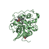
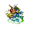
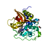
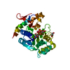
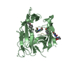
- PDB-4i07: Structure of mature form of cathepsin B1 from Schistosoma mansoni -
+ Open data

- Basic information
| Entry | Database: PDB / ID: 4i07 | ||||||
|---|---|---|---|---|---|---|---|
| Title | Structure of mature form of cathepsin B1 from Schistosoma mansoni | ||||||
 Components Components | Cathepsin B-like peptidase (C01 family) | ||||||
 Keywords Keywords |  HYDROLASE / peptidase / digestive tract HYDROLASE / peptidase / digestive tract | ||||||
| Function / homology |  Function and homology information Function and homology information | ||||||
| Biological species |  Schistosoma mansoni (invertebrata) Schistosoma mansoni (invertebrata) | ||||||
| Method | X-RAY DIFFRACTION / SYNCHROTRON / MOLECULAR REPLACEMENT / Resolution: 1.3 Å | ||||||
 Authors Authors | Rezacova, P. / Jilkova, A. / Brynda, J. / Horn, M. / Mares, M. | ||||||
 Citation Citation | Journal: Structure / Year: 2014 Title: Activation route of the Schistosoma mansoni cathepsin B1 drug target: structural map with a glycosaminoglycan switch Authors: Jilkova, A. / Horn, M. / Rezacova, P. / Maresova, L. / Fajtova, P. / Brynda, J. / Vondrasek, J. / McKerrow, J.H. / Caffrey, C.R. / Mares, M. | ||||||
| History |
|
- Structure visualization
| Structure viewer | Molecule: MolmilJmol/JSmol |
|---|
- Downloads & links
-Download
| PDBx/mmCIF format | 4i07.cif.gz | 134.7 KB | Display | PDBx/mmCIF format |
|---|---|---|---|---|
| PDB format | pdb4i07.ent.gz | 103 KB | Display | PDB format |
| PDBx/mmJSON format | 4i07.json.gz | Tree view | PDBx/mmJSON format | |
| Others |  Other downloads Other downloads |
-Validation report
| Arichive directory | https://data.pdbj.org/pub/pdb/validation_reports/i0/4i07ftp://data.pdbj.org/pub/pdb/validation_reports/i0/4i07 | HTTPS FTP |
|---|
-Related structure data
| Related structure data | 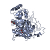 4i04C 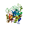 4i05C  3qsdS S: Starting model for refinement C: citing same article ( |
|---|---|
| Similar structure data |


-Links
 PDBj
PDBj
- Assembly
| Deposited unit | 
| ||||||||
|---|---|---|---|---|---|---|---|---|---|
| 1 |
| ||||||||
| Unit cell |
|
-Components
| #1: Protein | Mass: 28539.256 Da / Num. of mol.: 1 / Fragment: UNP residues 87-340 / Mutation: T168A, T283A Source method: isolated from a genetically manipulated source Details: zeocin resistance / Source: (gene. exp.) Schistosoma mansoni (invertebrata) / Gene: cb1.1, Smp_103610 / Plasmid: pPICZalpha / Production host:  Pichia pastoris (fungus) / Strain (production host): X33 / References: UniProt: Q8MNY2, cathepsin B Pichia pastoris (fungus) / Strain (production host): X33 / References: UniProt: Q8MNY2, cathepsin B |
|---|---|
| #2: Chemical | ChemComp-CL / Chloride  Mass: 35.453 Da / Num. of mol.: 1 / Source method: obtained synthetically / Formula: Cl Mass: 35.453 Da / Num. of mol.: 1 / Source method: obtained synthetically / Formula: Cl |
| #3: Chemical | ChemComp-ACT / Acetate  Mass: 59.044 Da / Num. of mol.: 1 / Source method: obtained synthetically / Formula: C2H3O2 Mass: 59.044 Da / Num. of mol.: 1 / Source method: obtained synthetically / Formula: C2H3O2 |
| #4: Water | ChemComp-HOH / Water Mass: 18.015 Da / Num. of mol.: 427 / Source method: isolated from a natural source / Formula: H2O Mass: 18.015 Da / Num. of mol.: 427 / Source method: isolated from a natural source / Formula: H2O |
-Experimental details
-Experiment
| Experiment | Method: X-RAY DIFFRACTION / Number of used crystals: 1 |
|---|
- Sample preparation
| Crystal | Density Matthews: 2.08 Å3/Da / Density % sol: 40.86 % |
|---|---|
| Crystal grow | Temperature: 293 K / Method: vapor diffusion, hanging drop / pH: 5.6 Details: Reservoir: 0.2M Ammonium Acetate, 0.1M Sodium Citrate, 30% PEG 4000. Protein buffer and concentration: 5mM Sodium Acetate, pH 5.5, Cpr=2.5mg/ml. Ratio Protein: Reservoir=1:1. Cryocooled in ...Details: Reservoir: 0.2M Ammonium Acetate, 0.1M Sodium Citrate, 30% PEG 4000. Protein buffer and concentration: 5mM Sodium Acetate, pH 5.5, Cpr=2.5mg/ml. Ratio Protein: Reservoir=1:1. Cryocooled in mother liquor, VAPOR DIFFUSION, HANGING DROP, temperature 293K |
-Data collection
| Diffraction | Mean temperature: 100 K |
|---|---|
| Diffraction source | Source: SYNCHROTRON / Site: APS  / Beamline: 19-BM / Wavelength: 0.978 Å / Beamline: 19-BM / Wavelength: 0.978 Å |
| Detector | Type: ADSC QUANTUM 210r / Detector: CCD / Date: Jul 10, 2008 / Details: mirrors |
| Radiation | Monochromator: Si111 double-crystal monochromator / Protocol: SINGLE WAVELENGTH / Monochromatic (M) / Laue (L): M / Scattering type: x-ray |
| Radiation wavelength | Wavelength: 0.978 Å / Relative weight: 1 |
| Reflection | Resolution: 1.3→50 Å / Num. all: 66361 / Num. obs: 59658 / % possible obs: 89.9 % / Observed criterion σ(F): 0 / Observed criterion σ(I): 0 / Redundancy: 7.3 % / Biso Wilson estimate: 13.5 Å2 / Rmerge(I) obs: 0.077 / Net I/σ(I): 33.7 |
| Reflection shell | Resolution: 1.3→1.32 Å / Redundancy: 5.7 % / Rmerge(I) obs: 0.438 / Mean I/σ(I) obs: 4 / % possible all: 49.3 |
- Processing
| Software |
| ||||||||||||||||||||||||||||||||||||||||||||||||||||||||||||||||||||||||||||||||||||||||||||||||||||||||||||||||||||||||||||||||||||||||||||||||||||||||||||||||||||||||||
|---|---|---|---|---|---|---|---|---|---|---|---|---|---|---|---|---|---|---|---|---|---|---|---|---|---|---|---|---|---|---|---|---|---|---|---|---|---|---|---|---|---|---|---|---|---|---|---|---|---|---|---|---|---|---|---|---|---|---|---|---|---|---|---|---|---|---|---|---|---|---|---|---|---|---|---|---|---|---|---|---|---|---|---|---|---|---|---|---|---|---|---|---|---|---|---|---|---|---|---|---|---|---|---|---|---|---|---|---|---|---|---|---|---|---|---|---|---|---|---|---|---|---|---|---|---|---|---|---|---|---|---|---|---|---|---|---|---|---|---|---|---|---|---|---|---|---|---|---|---|---|---|---|---|---|---|---|---|---|---|---|---|---|---|---|---|---|---|---|---|---|---|
| Refinement | Method to determine structure: MOLECULAR REPLACEMENT Starting model: 3QSD Resolution: 1.3→19.79 Å / Cor.coef. Fo:Fc: 0.973 / Cor.coef. Fo:Fc free: 0.959 / SU B: 1.487 / SU ML: 0.029 / Cross valid method: THROUGHOUT / σ(F): 0 / σ(I): 0 / ESU R: 0.057 / ESU R Free: 0.054 / Stereochemistry target values: MAXIMUM LIKELIHOOD / Details: HYDROGENS HAVE BEEN ADDED IN THE RIDING POSITIONS
| ||||||||||||||||||||||||||||||||||||||||||||||||||||||||||||||||||||||||||||||||||||||||||||||||||||||||||||||||||||||||||||||||||||||||||||||||||||||||||||||||||||||||||
| Solvent computation | Ion probe radii: 0.8 Å / Shrinkage radii: 0.8 Å / VDW probe radii: 1.2 Å / Solvent model: BABINET MODEL WITH MASK | ||||||||||||||||||||||||||||||||||||||||||||||||||||||||||||||||||||||||||||||||||||||||||||||||||||||||||||||||||||||||||||||||||||||||||||||||||||||||||||||||||||||||||
| Displacement parameters | Biso mean: 11.512 Å2
| ||||||||||||||||||||||||||||||||||||||||||||||||||||||||||||||||||||||||||||||||||||||||||||||||||||||||||||||||||||||||||||||||||||||||||||||||||||||||||||||||||||||||||
| Refinement step | Cycle: LAST / Resolution: 1.3→19.79 Å
| ||||||||||||||||||||||||||||||||||||||||||||||||||||||||||||||||||||||||||||||||||||||||||||||||||||||||||||||||||||||||||||||||||||||||||||||||||||||||||||||||||||||||||
| Refine LS restraints |
| ||||||||||||||||||||||||||||||||||||||||||||||||||||||||||||||||||||||||||||||||||||||||||||||||||||||||||||||||||||||||||||||||||||||||||||||||||||||||||||||||||||||||||
| LS refinement shell | Resolution: 1.3→1.334 Å / Total num. of bins used: 20
|
