 Movie
Movie Controller

[English] 日本語
 Yorodumi
- PDB-4hvp: Structure of complex of synthetic HIV-1 protease with a substrate... -
+ Open data

- Basic information
| Entry | Database: PDB / ID: 4hvp | ||||||
|---|---|---|---|---|---|---|---|
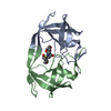
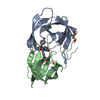
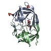
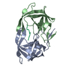
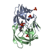
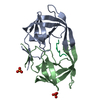
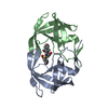
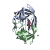
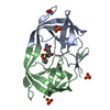
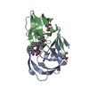
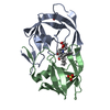
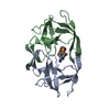
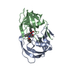
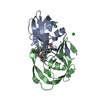
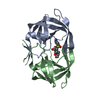
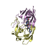
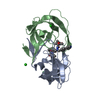
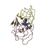
| Title | Structure of complex of synthetic HIV-1 protease with a substrate-based inhibitor at 2.3 Angstroms resolution | ||||||
 Components Components | HIV-1 PROTEASE | ||||||
 Keywords Keywords | HYDROLASE/HYDROLASE INHIBITOR / ACID PROTEINASE / HYDROLASE-HYDROLASE INHIBITOR complex | ||||||
| Function / homology |  Function and homology information Function and homology informationRNA stem-loop binding / host cell / HIV-1 retropepsin / retroviral ribonuclease H / exoribonuclease H / exoribonuclease H activity / host multivesicular body / DNA integration / RNA-directed DNA polymerase / viral genome integration into host DNA ...RNA stem-loop binding / host cell / HIV-1 retropepsin / retroviral ribonuclease H / exoribonuclease H / exoribonuclease H activity / host multivesicular body / DNA integration / RNA-directed DNA polymerase / viral genome integration into host DNA / viral penetration into host nucleus / establishment of integrated proviral latency / RNA-directed DNA polymerase activity / RNA-DNA hybrid ribonuclease activity / Transferases; Transferring phosphorus-containing groups; Nucleotidyltransferases / symbiont-mediated suppression of host gene expression / viral nucleocapsid / DNA recombination / Hydrolases; Acting on ester bonds / DNA-directed DNA polymerase / aspartic-type endopeptidase activity / DNA-directed DNA polymerase activity / symbiont entry into host cell / lipid binding / host cell nucleus / host cell plasma membrane / virion membrane / structural molecule activity / proteolysis / DNA binding / zinc ion binding / membraneSimilarity search - Function | ||||||
| Biological species |   Human immunodeficiency virus 1 Human immunodeficiency virus 1 | ||||||
| Method | X-RAY DIFFRACTION / Resolution: 2.3 Å | ||||||
 Authors Authors | Miller, M. / Schneider, J. / Sathyanarayana, B.K. / Toth, M.V. / Marshall, G.R. / Clawson, L. / Selk, L. / Kent, S.B.H. / Wlodawer, A. | ||||||
 Citation Citation | Journal: Science / Year: 1989 Title: Structure of complex of synthetic HIV-1 protease with a substrate-based inhibitor at 2.3 A resolution. Authors: Miller, M. / Schneider, J. / Sathyanarayana, B.K. / Toth, M.V. / Marshall, G.R. / Clawson, L. / Selk, L. / Kent, S.B. / Wlodawer, A. #1: Journal: Science / Year: 1989Title: Conserved Folding in Retroviral Proteases. Crystal Structure of a Synthetic HIV-1 Protease Authors: Wlodawer, A. / Miller, M. / Jaskolski, M. / Sathyanarayana, B.K. / Baldwin, E. / Weber, I.T. / Selk, L.M. / Clawson, L. / Schneider, J. / Kent, S.B.H. #2: Journal: Science / Year: 1989Title: Molecular Modeling of the HIV-1 Protease and its Substrate Binding Site Authors: Weber, I.T. / Miller, M. / Jaskolski, M. / Leis, J. / Skalka, A.M. / Wlodawer, A. #3: Journal: Nature / Year: 1989Title: Crystal Structure of a Retroviral Protease Proves Relationship to Aspartic Protease Family Authors: Miller, M. / Jaskolski, M. / Rao, J.K.M. / Leis, J. / Wlodawer, A. #4: Journal: Cell(Cambridge,Mass.) / Year: 1988Title: Enzymatic Activity of a Synthetic 99 Residue Protein Corresponding to the Putative HIV-1 Protease Authors: Schneider, J. / Kent, S.B.H. | ||||||
| History |
| ||||||
| Remark 700 | THE DIMER INTERFACE IS COMPOSED OF INTERDIGITATED N- AND C-TERMINI FROM BOTH SUBUNITS FORMING A ...THE DIMER INTERFACE IS COMPOSED OF INTERDIGITATED N- AND C-TERMINI FROM BOTH SUBUNITS FORMING A FOUR-STRANDED ANTIPARALLEL BETA-SHEET. |
- Structure visualization
| Structure viewer | Molecule: MolmilJmol/JSmol |
|---|
- Downloads & links
-Download
| PDBx/mmCIF format | 4hvp.cif.gz | 51.1 KB | Display | PDBx/mmCIF format |
|---|---|---|---|---|
| PDB format | pdb4hvp.ent.gz | 39.3 KB | Display | PDB format |
| PDBx/mmJSON format | 4hvp.json.gz | Tree view | PDBx/mmJSON format | |
| Others |  Other downloads Other downloads |
-Validation report
| Arichive directory | https://data.pdbj.org/pub/pdb/validation_reports/hv/4hvpftp://data.pdbj.org/pub/pdb/validation_reports/hv/4hvp | HTTPS FTP |
|---|
-Related structure data
| Similar structure data |
|---|


-Links
 PDBj
PDBj

- Assembly
| Deposited unit | 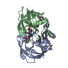
| ||||||||
|---|---|---|---|---|---|---|---|---|---|
| 1 |
| ||||||||
| Unit cell |
| ||||||||
| Atom site foot note | 1: THE PEPTIDE BOND BETWEEN RESIDUES NLE I 3 AND NLE I 4 HAS BEEN REDUCED AND IS -CH2-NH-. |
-Components
| #1: Protein | Mass: 10764.636 Da / Num. of mol.: 2 Source method: isolated from a genetically manipulated source Source: (gene. exp.) Human immunodeficiency virus 1 / Genus: Lentivirus / Production host:  Escherichia coli (E. coli) / References: UniProt: P03369 Escherichia coli (E. coli) / References: UniProt: P03369#2: Chemical |   Type: peptide-like, Peptide-like / Class: Inhibitor / Mass: 770.983 Da / Num. of mol.: 1 / Source method: obtained synthetically / Formula: C35H68N11O8 Type: peptide-like, Peptide-like / Class: Inhibitor / Mass: 770.983 Da / Num. of mol.: 1 / Source method: obtained synthetically / Formula: C35H68N11O8References: N-{(2S)-2-[(N-acetyl-L-threonyl-L-isoleucyl)amino]hexyl}-L-norleucyl-L-glutaminyl-N~5~-[amino(iminio)methyl]-L-ornithinamide #3: Water | ChemComp-HOH / | Water Mass: 18.015 Da / Num. of mol.: 70 / Source method: isolated from a natural source / Formula: H2O Mass: 18.015 Da / Num. of mol.: 70 / Source method: isolated from a natural source / Formula: H2ONonpolymer details | THE PEPTIDE BOND BETWEEN SUBCOMPONE | |
|---|
-Experimental details
-Experiment
| Experiment | Method: X-RAY DIFFRACTION |
|---|
- Sample preparation
| Crystal | Density Matthews: 2.14 Å3/Da / Density % sol: 42.57 % | |||||||||||||||
|---|---|---|---|---|---|---|---|---|---|---|---|---|---|---|---|---|
| Crystal grow | *PLUS Method: vapor diffusion, hanging drop / pH: 5.4 | |||||||||||||||
| Components of the solutions | *PLUS
|
-Data collection
| Radiation | Scattering type: x-ray |
|---|---|
| Radiation wavelength | Relative weight: 1 |
| Reflection | *PLUS Highest resolution: 2.25 Å / Num. all: 8740 / Num. obs: 7943 / Observed criterion σ(I): 1.5 / Num. measured all: 55569 / Rmerge(I) obs: 0.068 |
- Processing
| Software | Name: PROLSQ / Classification: refinement | ||||||||||||||||||||||||||||||||||||||||||||||||||||||||||||||||||||||||||||||||||||
|---|---|---|---|---|---|---|---|---|---|---|---|---|---|---|---|---|---|---|---|---|---|---|---|---|---|---|---|---|---|---|---|---|---|---|---|---|---|---|---|---|---|---|---|---|---|---|---|---|---|---|---|---|---|---|---|---|---|---|---|---|---|---|---|---|---|---|---|---|---|---|---|---|---|---|---|---|---|---|---|---|---|---|---|---|---|
| Refinement | Resolution: 2.3→10 Å /
| ||||||||||||||||||||||||||||||||||||||||||||||||||||||||||||||||||||||||||||||||||||
| Refinement step | Cycle: LAST / Resolution: 2.3→10 Å
| ||||||||||||||||||||||||||||||||||||||||||||||||||||||||||||||||||||||||||||||||||||
| Refine LS restraints |
| ||||||||||||||||||||||||||||||||||||||||||||||||||||||||||||||||||||||||||||||||||||
| Software | *PLUS Name: 'X-PLOR, PROLSQ' / Classification: refinement | ||||||||||||||||||||||||||||||||||||||||||||||||||||||||||||||||||||||||||||||||||||
| Refinement | *PLUS Rfactor obs: 0.176 / Highest resolution: 2.25 Å | ||||||||||||||||||||||||||||||||||||||||||||||||||||||||||||||||||||||||||||||||||||
| Solvent computation | *PLUS | ||||||||||||||||||||||||||||||||||||||||||||||||||||||||||||||||||||||||||||||||||||
| Displacement parameters | *PLUS |
