 Movie
Movie Controller

[English] 日本語
 Yorodumi
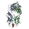
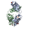
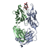
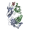
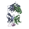
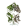
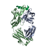
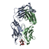
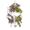
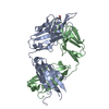
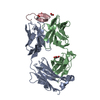
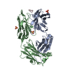
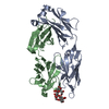
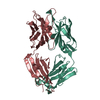
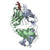
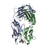
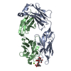
- PDB-4hs8: Hepatitus C envelope glycoprotein E2 fragment 412-423 with humani... -
+ Open data

- Basic information
| Entry | Database: PDB / ID: 4hs8 | ||||||
|---|---|---|---|---|---|---|---|
| Title | Hepatitus C envelope glycoprotein E2 fragment 412-423 with humanized and affinity-matured antibody hu5B3.v3 | ||||||
 Components Components |
| ||||||
 Keywords Keywords |  IMMUNE SYSTEM IMMUNE SYSTEM | ||||||
| Function / homology |  Function and homology information Function and homology informationhost cell lipid droplet / lipid droplet / host cell endoplasmic reticulum membrane / viral envelope / virion membrane / membraneSimilarity search - Function | ||||||
| Biological species |  Homo sapiens (human) Homo sapiens (human) Hepatitis C virus Hepatitis C virus | ||||||
| Method | X-RAY DIFFRACTION / SYNCHROTRON / MOLECULAR REPLACEMENT / Resolution: 2.6 Å | ||||||
 Authors Authors | Eigenbrot, C. / Ultsch, M. | ||||||
 Citation Citation | Journal: J.Mol.Biol. / Year: 2013 Title: Glycan shifting on hepatitis C virus (HCV) e2 glycoprotein is a mechanism for escape from broadly neutralizing antibodies. Authors: Pantua, H. / Diao, J. / Ultsch, M. / Hazen, M. / Mathieu, M. / McCutcheon, K. / Takeda, K. / Date, S. / Cheung, T.K. / Phung, Q. / Hass, P. / Arnott, D. / Hongo, J.A. / Matthews, D.J. / ...Authors: Pantua, H. / Diao, J. / Ultsch, M. / Hazen, M. / Mathieu, M. / McCutcheon, K. / Takeda, K. / Date, S. / Cheung, T.K. / Phung, Q. / Hass, P. / Arnott, D. / Hongo, J.A. / Matthews, D.J. / Brown, A. / Patel, A.H. / Kelley, R.F. / Eigenbrot, C. / Kapadia, S.B. | ||||||
| History |
|
- Structure visualization
| Structure viewer | Molecule: MolmilJmol/JSmol |
|---|
- Downloads & links
-Download
| PDBx/mmCIF format | 4hs8.cif.gz | 191.1 KB | Display | PDBx/mmCIF format |
|---|---|---|---|---|
| PDB format | pdb4hs8.ent.gz | 151.7 KB | Display | PDB format |
| PDBx/mmJSON format | 4hs8.json.gz | Tree view | PDBx/mmJSON format | |
| Others |  Other downloads Other downloads |
-Validation report
| Arichive directory | https://data.pdbj.org/pub/pdb/validation_reports/hs/4hs8ftp://data.pdbj.org/pub/pdb/validation_reports/hs/4hs8 | HTTPS FTP |
|---|
-Related structure data
| Related structure data | 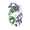 4hs6C  1fvcS C: citing same article ( S: Starting model for refinement |
|---|---|
| Similar structure data |


-Links
 PDBj
PDBj


- Assembly
| Deposited unit | 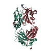
| ||||||||
|---|---|---|---|---|---|---|---|---|---|
| 1 |
| ||||||||
| Unit cell |
|
-Components
-Protein/peptide , 1 types, 1 molecules A
| #1: Protein/peptide | Mass: 1727.877 Da / Num. of mol.: 1 / Fragment: UNP residues 50-62 / Source method: obtained synthetically Details: This sequence occurs naturally in hepatitus C virus Source: (synth.) Hepatitis C virus / References: UniProt: Q9YK84 |
|---|
-Antibody , 2 types, 2 molecules HL
| #2: Antibody | Mass: 24653.465 Da / Num. of mol.: 1 Source method: isolated from a genetically manipulated source Source: (gene. exp.) Homo sapiens (human) / Production host:  Escherichia coli (E. coli) Escherichia coli (E. coli) |
|---|---|
| #3: Antibody | Mass: 23767.291 Da / Num. of mol.: 1 Source method: isolated from a genetically manipulated source Source: (gene. exp.) Homo sapiens (human) / Production host: Escherichia coli (E. coli) |
-Non-polymers , 3 types, 31 molecules 


| #4: Chemical | Glycerol Mass: 92.094 Da / Num. of mol.: 3 / Source method: obtained synthetically / Formula: C3H8O3 Mass: 92.094 Da / Num. of mol.: 3 / Source method: obtained synthetically / Formula: C3H8O3#5: Chemical | Sulfate Mass: 96.063 Da / Num. of mol.: 2 / Source method: obtained synthetically / Formula: SO4 Mass: 96.063 Da / Num. of mol.: 2 / Source method: obtained synthetically / Formula: SO4#6: Water | ChemComp-HOH / | WaterMass: 18.015 Da / Num. of mol.: 26 / Source method: isolated from a natural source / Formula: H2O |
|---|
-Details
| Nonpolymer details | E2-PEPTIDE IS BIOTINYLATED, WITH BIOTIN (BTN) ATTACHED TO THE SIDE CHAIN OF THE C-TERMINAL LYS ...E2-PEPTIDE IS BIOTINYLAT |
|---|
-Experimental details
-Experiment
| Experiment | Method: X-RAY DIFFRACTION / Number of used crystals: 1 |
|---|
- Sample preparation
| Crystal | Density Matthews: 2.73 Å3/Da / Density % sol: 54.96 % |
|---|---|
| Crystal grow | Temperature: 291 K / Method: vapor diffusion, sitting drop / pH: 6 Details: ammonium sulfate, pH 6.0, VAPOR DIFFUSION, SITTING DROP, temperature 291K |
-Data collection
| Diffraction | Mean temperature: 110 K |
|---|---|
| Diffraction source | Source: SYNCHROTRON / Site: SSRL  / Beamline: BL11-1 / Wavelength: 0.98 Å / Beamline: BL11-1 / Wavelength: 0.98 Å |
| Detector | Type: MARMOSAIC 325 mm CCD / Detector: CCD / Date: Mar 16, 2011 |
| Radiation | Monochromator: Si (111) / Protocol: SINGLE WAVELENGTH / Monochromatic (M) / Laue (L): M / Scattering type: x-ray |
| Radiation wavelength | Wavelength: 0.98 Å / Relative weight: 1 |
| Reflection | Resolution: 2.6→50 Å / Num. all: 17898 / Num. obs: 17880 / % possible obs: 99.9 % / Observed criterion σ(I): -3 / Redundancy: 7.7 % / Biso Wilson estimate: 73.69 Å2 / Rsym value: 0.098 / Net I/σ(I): 19 |
- Processing
| Software |
| ||||||||||||||||||||||||||||||||||||||||||||||||||||||||||||||||||||||||||||||||||||||||||||||||||||||||||||||||||||||||||||||||||||||||||||||||||||||
|---|---|---|---|---|---|---|---|---|---|---|---|---|---|---|---|---|---|---|---|---|---|---|---|---|---|---|---|---|---|---|---|---|---|---|---|---|---|---|---|---|---|---|---|---|---|---|---|---|---|---|---|---|---|---|---|---|---|---|---|---|---|---|---|---|---|---|---|---|---|---|---|---|---|---|---|---|---|---|---|---|---|---|---|---|---|---|---|---|---|---|---|---|---|---|---|---|---|---|---|---|---|---|---|---|---|---|---|---|---|---|---|---|---|---|---|---|---|---|---|---|---|---|---|---|---|---|---|---|---|---|---|---|---|---|---|---|---|---|---|---|---|---|---|---|---|---|---|---|---|---|---|
| Refinement | Method to determine structure: MOLECULAR REPLACEMENT Starting model: 1FVC Resolution: 2.6→29.51 Å / Cor.coef. Fo:Fc: 0.9298 / Cor.coef. Fo:Fc free: 0.907 / SU R Cruickshank DPI: 0.564 / Isotropic thermal model: TLS in 5 groups / Cross valid method: THROUGHOUT / σ(F): 0 / Stereochemistry target values: maXIMUM LIKELIHOOD
| ||||||||||||||||||||||||||||||||||||||||||||||||||||||||||||||||||||||||||||||||||||||||||||||||||||||||||||||||||||||||||||||||||||||||||||||||||||||
| Displacement parameters | Biso mean: 65.1 Å2
| ||||||||||||||||||||||||||||||||||||||||||||||||||||||||||||||||||||||||||||||||||||||||||||||||||||||||||||||||||||||||||||||||||||||||||||||||||||||
| Refine analyze | Luzzati coordinate error obs: 0.441 Å | ||||||||||||||||||||||||||||||||||||||||||||||||||||||||||||||||||||||||||||||||||||||||||||||||||||||||||||||||||||||||||||||||||||||||||||||||||||||
| Refinement step | Cycle: LAST / Resolution: 2.6→29.51 Å
| ||||||||||||||||||||||||||||||||||||||||||||||||||||||||||||||||||||||||||||||||||||||||||||||||||||||||||||||||||||||||||||||||||||||||||||||||||||||
| Refine LS restraints |
| ||||||||||||||||||||||||||||||||||||||||||||||||||||||||||||||||||||||||||||||||||||||||||||||||||||||||||||||||||||||||||||||||||||||||||||||||||||||
| LS refinement shell | Resolution: 2.6→2.76 Å / Total num. of bins used: 9
| ||||||||||||||||||||||||||||||||||||||||||||||||||||||||||||||||||||||||||||||||||||||||||||||||||||||||||||||||||||||||||||||||||||||||||||||||||||||
| Refinement TLS params. | Method: refined / Refine-ID: X-RAY DIFFRACTION
| ||||||||||||||||||||||||||||||||||||||||||||||||||||||||||||||||||||||||||||||||||||||||||||||||||||||||||||||||||||||||||||||||||||||||||||||||||||||
| Refinement TLS group |
|
