 Movie
Movie Controller


+ Open data

- Basic information
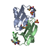
| Entry | Database: PDB / ID: 4hs2 | ||||||
|---|---|---|---|---|---|---|---|
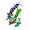
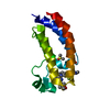
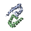
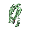
| Title | Crystal Structure of the Human SPOP C-terminal Domain | ||||||
 Components Components | Speckle-type POZ protein | ||||||
 Keywords Keywords |  PROTEIN BINDING / protein interaction domain / Oligomerisation PROTEIN BINDING / protein interaction domain / Oligomerisation | ||||||
| Function / homology |  Function and homology informationregulation of proteolysis / Cul3-RING ubiquitin ligase complex / molecular function inhibitor activity / localization / Hedgehog 'on' state / protein polyubiquitination / proteasome-mediated ubiquitin-dependent protein catabolic process / nuclear speck / ubiquitin protein ligase binding / nucleoplasm ...regulation of proteolysis / Cul3-RING ubiquitin ligase complex / molecular function inhibitor activity / localization / Hedgehog 'on' state / protein polyubiquitination / proteasome-mediated ubiquitin-dependent protein catabolic process / nuclear speck / ubiquitin protein ligase binding / nucleoplasm / nucleus / cytoplasm Function and homology informationregulation of proteolysis / Cul3-RING ubiquitin ligase complex / molecular function inhibitor activity / localization / Hedgehog 'on' state / protein polyubiquitination / proteasome-mediated ubiquitin-dependent protein catabolic process / nuclear speck / ubiquitin protein ligase binding / nucleoplasm ...regulation of proteolysis / Cul3-RING ubiquitin ligase complex / molecular function inhibitor activity / localization / Hedgehog 'on' state / protein polyubiquitination / proteasome-mediated ubiquitin-dependent protein catabolic process / nuclear speck / ubiquitin protein ligase binding / nucleoplasm / nucleus / cytoplasmSimilarity search - Function | ||||||
| Biological species |  Homo sapiens (human) Homo sapiens (human) | ||||||
| Method | X-RAY DIFFRACTION / SYNCHROTRON / SAD / Resolution: 1.53 Å | ||||||
 Authors Authors | Van Geersdaele, L.K. / Stead, M.A. / Carr, S.B. / Wright, S.C. | ||||||
 Citation Citation | Journal: Acta Crystallogr.,Sect.D / Year: 2013 Title: Structural basis of high-order oligomerization of the cullin-3 adaptor SPOP. Authors: van Geersdaele, L.K. / Stead, M.A. / Harrison, C.M. / Carr, S.B. / Close, H.J. / Rosbrook, G.O. / Connell, S.D. / Wright, S.C. | ||||||
| History |
|
- Structure visualization
| Structure viewer | Molecule: MolmilJmol/JSmol |
|---|
- Downloads & links
-Download
| PDBx/mmCIF format | 4hs2.cif.gz | 43.4 KB | Display | PDBx/mmCIF format |
|---|---|---|---|---|
| PDB format | pdb4hs2.ent.gz | 29.8 KB | Display | PDB format |
| PDBx/mmJSON format | 4hs2.json.gz | Tree view | PDBx/mmJSON format | |
| Others |  Other downloads Other downloads |
-Validation report
| Arichive directory | https://data.pdbj.org/pub/pdb/validation_reports/hs/4hs2ftp://data.pdbj.org/pub/pdb/validation_reports/hs/4hs2 | HTTPS FTP |
|---|
-Related structure data


-Links
 PDBj
PDBj


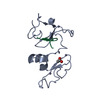
- Assembly
| Deposited unit | 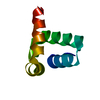
| ||||||||
|---|---|---|---|---|---|---|---|---|---|
| 1 | 
| ||||||||
| Unit cell |
| ||||||||
| Components on special symmetry positions |
|
-Components
| #1: Protein | Mass: 11941.446 Da / Num. of mol.: 1 / Fragment: C-terminal domain (UNP residues 270-374) / Mutation: L273D, L282D, L285K Source method: isolated from a genetically manipulated source Source: (gene. exp.) Homo sapiens (human) / Gene: SPOP / Plasmid: pGEX-6P-1 / Production host:  Escherichia coli (E. coli) / Strain (production host): BL21(DE3)pLysS; B834 / References: UniProt: O43791 Escherichia coli (E. coli) / Strain (production host): BL21(DE3)pLysS; B834 / References: UniProt: O43791 |
|---|---|
| #2: Water | ChemComp-HOH / Water Mass: 18.015 Da / Num. of mol.: 62 / Source method: isolated from a natural source / Formula: H2O Mass: 18.015 Da / Num. of mol.: 62 / Source method: isolated from a natural source / Formula: H2O |
-Experimental details
-Experiment
| Experiment | Method: X-RAY DIFFRACTION / Number of used crystals: 1 |
|---|
- Sample preparation
| Crystal | Density Matthews: 2.07 Å3/Da / Density % sol: 40.44 % |
|---|---|
| Crystal grow | Temperature: 291 K / Method: vapor diffusion, hanging drop / pH: 6.5 Details: 26% PEG 1500, 7% isopropanol, 0.1M CaCl2, 0.1M imidazole pH 6.5, VAPOR DIFFUSION, HANGING DROP, temperature 291K |
-Data collection
| Diffraction |
| ||||||||||||||||||||||||||||||||||||||||||||||||||||||||||||||||||||||||||||||||||||||||||||||||||||||||||||||||||||||||||||||||||||||||||||||||||||||||||||||||||||||||
|---|---|---|---|---|---|---|---|---|---|---|---|---|---|---|---|---|---|---|---|---|---|---|---|---|---|---|---|---|---|---|---|---|---|---|---|---|---|---|---|---|---|---|---|---|---|---|---|---|---|---|---|---|---|---|---|---|---|---|---|---|---|---|---|---|---|---|---|---|---|---|---|---|---|---|---|---|---|---|---|---|---|---|---|---|---|---|---|---|---|---|---|---|---|---|---|---|---|---|---|---|---|---|---|---|---|---|---|---|---|---|---|---|---|---|---|---|---|---|---|---|---|---|---|---|---|---|---|---|---|---|---|---|---|---|---|---|---|---|---|---|---|---|---|---|---|---|---|---|---|---|---|---|---|---|---|---|---|---|---|---|---|---|---|---|---|---|---|---|---|
| Diffraction source |
| ||||||||||||||||||||||||||||||||||||||||||||||||||||||||||||||||||||||||||||||||||||||||||||||||||||||||||||||||||||||||||||||||||||||||||||||||||||||||||||||||||||||||
| Detector | Type: PSI PILATUS 6M / Detector: PIXEL / Date: Jul 15, 2011 | ||||||||||||||||||||||||||||||||||||||||||||||||||||||||||||||||||||||||||||||||||||||||||||||||||||||||||||||||||||||||||||||||||||||||||||||||||||||||||||||||||||||||
| Radiation |
| ||||||||||||||||||||||||||||||||||||||||||||||||||||||||||||||||||||||||||||||||||||||||||||||||||||||||||||||||||||||||||||||||||||||||||||||||||||||||||||||||||||||||
| Radiation wavelength |
| ||||||||||||||||||||||||||||||||||||||||||||||||||||||||||||||||||||||||||||||||||||||||||||||||||||||||||||||||||||||||||||||||||||||||||||||||||||||||||||||||||||||||
| Reflection | Redundancy: 71.5 % / Av σ(I) over netI: 3.4 / Number: 442290 / Rsym value: 0.185 / D res high: 2.125 Å / D res low: 51.405 Å / Num. obs: 6183 / % possible obs: 100 | ||||||||||||||||||||||||||||||||||||||||||||||||||||||||||||||||||||||||||||||||||||||||||||||||||||||||||||||||||||||||||||||||||||||||||||||||||||||||||||||||||||||||
| Diffraction reflection shell |
| ||||||||||||||||||||||||||||||||||||||||||||||||||||||||||||||||||||||||||||||||||||||||||||||||||||||||||||||||||||||||||||||||||||||||||||||||||||||||||||||||||||||||
| Reflection | Resolution: 1.528→51.405 Å / Num. all: 16004 / Num. obs: 16004 / % possible obs: 99.8 % / Redundancy: 11.6 % / Rsym value: 0.056 / Net I/σ(I): 24.2 | ||||||||||||||||||||||||||||||||||||||||||||||||||||||||||||||||||||||||||||||||||||||||||||||||||||||||||||||||||||||||||||||||||||||||||||||||||||||||||||||||||||||||
| Reflection shell | Diffraction-ID: 1,2
|

-Phasing
| Phasing | Method: SAD |
|---|
- Processing
| Software |
| |||||||||||||||||||||||||||||||||||||||||||||||||||||||||||||||||||||||||||
|---|---|---|---|---|---|---|---|---|---|---|---|---|---|---|---|---|---|---|---|---|---|---|---|---|---|---|---|---|---|---|---|---|---|---|---|---|---|---|---|---|---|---|---|---|---|---|---|---|---|---|---|---|---|---|---|---|---|---|---|---|---|---|---|---|---|---|---|---|---|---|---|---|---|---|---|---|
| Refinement | Method to determine structure: SAD / Resolution: 1.53→27.75 Å / Cor.coef. Fo:Fc: 0.976 / Cor.coef. Fo:Fc free: 0.975 / WRfactor Rfree: 0.1444 / WRfactor Rwork: 0.1267 / Occupancy max: 1 / Occupancy min: 0.5 / FOM work R set: 0.9316 / SU B: 1.61 / SU ML: 0.027 / SU R Cruickshank DPI: 0.0512 / SU Rfree: 0.0469 / Cross valid method: THROUGHOUT / σ(F): 0 / ESU R: 0.051 / ESU R Free: 0.047 / Stereochemistry target values: MAXIMUM LIKELIHOOD Details: HYDROGENS HAVE BEEN ADDED IN THE RIDING POSITIONS. U VALUES: REFINED INDIVIDUALLY
| |||||||||||||||||||||||||||||||||||||||||||||||||||||||||||||||||||||||||||
| Solvent computation | Ion probe radii: 0.8 Å / Shrinkage radii: 0.8 Å / VDW probe radii: 1.2 Å / Solvent model: MASK | |||||||||||||||||||||||||||||||||||||||||||||||||||||||||||||||||||||||||||
| Displacement parameters | Biso max: 123.41 Å2 / Biso mean: 21.2669 Å2 / Biso min: 11.07 Å2
| |||||||||||||||||||||||||||||||||||||||||||||||||||||||||||||||||||||||||||
| Refinement step | Cycle: LAST / Resolution: 1.53→27.75 Å
| |||||||||||||||||||||||||||||||||||||||||||||||||||||||||||||||||||||||||||
| Refine LS restraints |
| |||||||||||||||||||||||||||||||||||||||||||||||||||||||||||||||||||||||||||
| LS refinement shell | Resolution: 1.528→1.567 Å / Total num. of bins used: 20
|
