 Movie
Movie Controller


+ Open data

- Basic information
| Entry | Database: PDB / ID: 4hex | ||||||
|---|---|---|---|---|---|---|---|
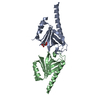
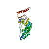
| Title | A novel conformation of calmodulin | ||||||
 Components Components | Calmodulin | ||||||
 Keywords Keywords | CALCIUM BINDING PROTEIN / Calmodulin / Novel conformation / trans / EF-hand motifs / Calcium signalling / Calcium binding / Neuromodulin / Neurogranin | ||||||
| Function / homology |  Function and homology information Function and homology informationCaMK IV-mediated phosphorylation of CREB / Cam-PDE 1 activation / CREB1 phosphorylation through the activation of CaMKII/CaMKK/CaMKIV cascasde / Glycogen breakdown (glycogenolysis) / Activation of RAC1 downstream of NMDARs / Reduction of cytosolic Ca++ levels / Sodium/Calcium exchangers / Activation of Ca-permeable Kainate Receptor / CLEC7A (Dectin-1) induces NFAT activation / Synthesis of IP3 and IP4 in the cytosol ...CaMK IV-mediated phosphorylation of CREB / Cam-PDE 1 activation / CREB1 phosphorylation through the activation of CaMKII/CaMKK/CaMKIV cascasde / Glycogen breakdown (glycogenolysis) / Activation of RAC1 downstream of NMDARs / Reduction of cytosolic Ca++ levels / Sodium/Calcium exchangers / Activation of Ca-permeable Kainate Receptor / CLEC7A (Dectin-1) induces NFAT activation / Synthesis of IP3 and IP4 in the cytosol / RHO GTPases activate PAKs / Calmodulin induced events / Inactivation, recovery and regulation of the phototransduction cascade / Tetrahydrobiopterin (BH4) synthesis, recycling, salvage and regulation / Calcineurin activates NFAT / eNOS activation / Ion transport by P-type ATPases / Unblocking of NMDA receptors, glutamate binding and activation / Protein methylation / RAF activation / VEGFR2 mediated vascular permeability / RAS processing / Smooth Muscle Contraction / Ca2+ pathway / negative regulation of calcium ion transmembrane transporter activity / FCERI mediated Ca+2 mobilization / RAF/MAP kinase cascade / RHO GTPases activate IQGAPs / Extra-nuclear estrogen signaling / PKA activation / regulation of response to tumor cell / positive regulation of autophagic cell death / DAPK1-calmodulin complex / Platelet degranulation / : / Stimuli-sensing channels / establishment of protein localization to mitochondrial membrane / Ion homeostasis / type 3 metabotropic glutamate receptor binding / regulation of synaptic vesicle endocytosis / negative regulation of high voltage-gated calcium channel activity / regulation of synaptic vesicle exocytosis / organelle localization by membrane tethering / negative regulation of calcium ion export across plasma membrane / mitochondrion-endoplasmic reticulum membrane tethering / regulation of cardiac muscle cell action potential / autophagosome membrane docking / response to corticosterone / positive regulation of ryanodine-sensitive calcium-release channel activity / nitric-oxide synthase binding / protein phosphatase activator activity / positive regulation of cyclic-nucleotide phosphodiesterase activity / positive regulation of phosphoprotein phosphatase activity / adenylate cyclase binding / catalytic complex / detection of calcium ion / negative regulation of ryanodine-sensitive calcium-release channel activity / regulation of cardiac muscle contraction / calcium channel inhibitor activity / cellular response to interferon-beta / phosphatidylinositol 3-kinase binding / enzyme regulator activity / regulation of release of sequestered calcium ion into cytosol by sarcoplasmic reticulum / regulation of calcium-mediated signaling / positive regulation of protein dephosphorylation / potassium ion transmembrane transport / voltage-gated potassium channel complex / regulation of ryanodine-sensitive calcium-release channel activity / titin binding / sperm midpiece / calcium channel complex / activation of adenylate cyclase activity / response to amphetamine / adenylate cyclase activator activity / regulation of heart rate / nitric-oxide synthase regulator activity / sarcomere / protein serine/threonine kinase activator activity / regulation of cytokinesis / calcium-mediated signaling / positive regulation of nitric-oxide synthase activity / mitochondrial membrane / positive regulation of receptor signaling pathway via JAK-STAT / spindle microtubule / synaptic vesicle membrane / cellular response to type II interferon / spindle pole / response to calcium ion / calcium-dependent protein binding / G2/M transition of mitotic cell cycle / disordered domain specific binding / myelin sheath / growth cone / vesicle / transmembrane transporter binding / protein autophosphorylation / positive regulation of apoptotic process / protein domain specific binding / centrosome / calcium ion bindingSimilarity search - Function | ||||||
| Biological species |  Mus musculus (house mouse) Mus musculus (house mouse) | ||||||
| Method | X-RAY DIFFRACTION / SYNCHROTRON / SAD / Resolution: 2.001 Å | ||||||
 Authors Authors | Kumar, V. / Chichili, V.P.R. / Sivaraman, J. | ||||||
 Citation Citation | Journal: Plos One / Year: 2013 Title: A novel trans conformation of ligand-free calmodulin Authors: Kumar, V. / Chichili, V.P.R. / Tang, X. / Sivaraman, J. | ||||||
| History |
|
- Structure visualization
| Structure viewer | Molecule: MolmilJmol/JSmol |
|---|
- Downloads & links
-Download
| PDBx/mmCIF format | 4hex.cif.gz | 126.4 KB | Display | PDBx/mmCIF format |
|---|---|---|---|---|
| PDB format | pdb4hex.ent.gz | 98.3 KB | Display | PDB format |
| PDBx/mmJSON format | 4hex.json.gz | Tree view | PDBx/mmJSON format | |
| Others |  Other downloads Other downloads |
-Validation report
| Arichive directory | https://data.pdbj.org/pub/pdb/validation_reports/he/4hexftp://data.pdbj.org/pub/pdb/validation_reports/he/4hex | HTTPS FTP |
|---|
-Related structure data
| Similar structure data |
|---|


-Links
 PDBj
PDBj


- Assembly
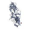
| Deposited unit | 
| ||||||||||||||||||
|---|---|---|---|---|---|---|---|---|---|---|---|---|---|---|---|---|---|---|---|
| 1 |
| ||||||||||||||||||
| Unit cell |
| ||||||||||||||||||
| Noncrystallographic symmetry (NCS) | NCS domain:
NCS domain segments: Component-ID: 0 / Ens-ID: 1 / Beg auth comp-ID: GLU / Beg label comp-ID: GLU / End auth comp-ID: VAL / End label comp-ID: VAL / Refine code: 0 / Auth seq-ID: 7 - 143 / Label seq-ID: 14 - 150
|
-Components
| #1: Protein | / CaM Mass: 17812.625 Da / Num. of mol.: 2 Source method: isolated from a genetically manipulated source Source: (gene. exp.) Mus musculus (house mouse) / Gene: Cam / Plasmid: pGS21a / Production host:  Escherichia coli (E. coli) / Strain (production host): BL21 (DE3) / References: UniProt: P62204, UniProt: P0DP26*PLUS Escherichia coli (E. coli) / Strain (production host): BL21 (DE3) / References: UniProt: P62204, UniProt: P0DP26*PLUS#2: Chemical | ChemComp-ZN /   Mass: 65.409 Da / Num. of mol.: 6 / Source method: obtained synthetically / Formula: Zn Mass: 65.409 Da / Num. of mol.: 6 / Source method: obtained synthetically / Formula: Zn#3: Chemical | ChemComp-CA /   Mass: 40.078 Da / Num. of mol.: 4 / Source method: obtained synthetically / Formula: Ca Mass: 40.078 Da / Num. of mol.: 4 / Source method: obtained synthetically / Formula: Ca#4: Water | ChemComp-HOH / | Water Mass: 18.015 Da / Num. of mol.: 78 / Source method: isolated from a natural source / Formula: H2O Mass: 18.015 Da / Num. of mol.: 78 / Source method: isolated from a natural source / Formula: H2O |
|---|
-Experimental details
-Experiment
| Experiment | Method: X-RAY DIFFRACTION / Number of used crystals: 1 |
|---|
- Sample preparation
| Crystal | Density Matthews: 2.45 Å3/Da / Density % sol: 49.84 % |
|---|---|
| Crystal grow | Temperature: 298 K / Method: vapor diffusion, hanging drop / pH: 8 Details: 100mM Tris-HCl, 8-10%(v/v) PEG 6K, 5-10mM ZnCl2, 10%(v/v) glycerol, pH 8.0, VAPOR DIFFUSION, HANGING DROP, temperature 298K |
-Data collection
| Diffraction | Mean temperature: 100 K | |||||||||||||||
|---|---|---|---|---|---|---|---|---|---|---|---|---|---|---|---|---|
| Diffraction source | Source: SYNCHROTRON / Site: NSLS  / Beamline: X8C / Wavelength: 0.978 Å / Beamline: X8C / Wavelength: 0.978 Å | |||||||||||||||
| Detector | Type: ADSC QUANTUM 4 / Detector: CCD / Date: Oct 25, 2009 | |||||||||||||||
| Radiation | Monochromator: Si 111 CHANNEL / Protocol: SINGLE WAVELENGTH / Monochromatic (M) / Laue (L): M / Scattering type: x-ray | |||||||||||||||
| Radiation wavelength | Wavelength: 0.978 Å / Relative weight: 1 | |||||||||||||||
| Reflection twin |
| |||||||||||||||
| Reflection | Resolution: 2.001→50 Å / Num. obs: 22800 / % possible obs: 99.3 % / Observed criterion σ(F): 0 / Observed criterion σ(I): 0 / Redundancy: 3.7 % / Rmerge(I) obs: 0.055 | |||||||||||||||
| Reflection shell | Resolution: 2.02→2.05 Å / Redundancy: 3.4 % / Rmerge(I) obs: 0.381 / Num. unique all: 2341 / % possible all: 98.9 |
- Processing
| Software |
| |||||||||||||||||||||||||||||||||||||||||||||||||||||||||||||||||||||||||||
|---|---|---|---|---|---|---|---|---|---|---|---|---|---|---|---|---|---|---|---|---|---|---|---|---|---|---|---|---|---|---|---|---|---|---|---|---|---|---|---|---|---|---|---|---|---|---|---|---|---|---|---|---|---|---|---|---|---|---|---|---|---|---|---|---|---|---|---|---|---|---|---|---|---|---|---|---|
| Refinement | Method to determine structure: SAD / Resolution: 2.001→28.58 Å / Cor.coef. Fo:Fc: 0.949 / Cor.coef. Fo:Fc free: 0.944 / Occupancy max: 1 / Occupancy min: 1 / SU B: 10.549 / SU ML: 0.163 / Cross valid method: THROUGHOUT / σ(F): 0 / ESU R: 0.039 / ESU R Free: 0.034 / Stereochemistry target values: MAXIMUM LIKELIHOOD Details: HYDROGENS HAVE BEEN ADDED IN THE RIDING POSITIONS U VALUES: RESIDUAL ONLY
| |||||||||||||||||||||||||||||||||||||||||||||||||||||||||||||||||||||||||||
| Solvent computation | Ion probe radii: 0.8 Å / Shrinkage radii: 0.8 Å / VDW probe radii: 1.2 Å / Solvent model: MASK | |||||||||||||||||||||||||||||||||||||||||||||||||||||||||||||||||||||||||||
| Displacement parameters | Biso max: 97.48 Å2 / Biso mean: 47.251 Å2 / Biso min: 25 Å2
| |||||||||||||||||||||||||||||||||||||||||||||||||||||||||||||||||||||||||||
| Refinement step | Cycle: LAST / Resolution: 2.001→28.58 Å
| |||||||||||||||||||||||||||||||||||||||||||||||||||||||||||||||||||||||||||
| Refine LS restraints |
| |||||||||||||||||||||||||||||||||||||||||||||||||||||||||||||||||||||||||||
| Refine LS restraints NCS | Ens-ID: 1 / Number: 6834 / Refine-ID: X-RAY DIFFRACTION / Type: interatomic distance / Rms dev position: 0.19 Å / Weight position: 0.05
| |||||||||||||||||||||||||||||||||||||||||||||||||||||||||||||||||||||||||||
| LS refinement shell | Resolution: 2.001→2.052 Å / Total num. of bins used: 20
| |||||||||||||||||||||||||||||||||||||||||||||||||||||||||||||||||||||||||||
| Refinement TLS params. | Method: refined / Refine-ID: X-RAY DIFFRACTION
| |||||||||||||||||||||||||||||||||||||||||||||||||||||||||||||||||||||||||||
| Refinement TLS group |
|
