 Movie
Movie Controller

[English] 日本語
 Yorodumi
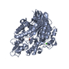
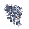
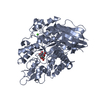
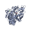
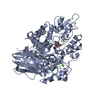
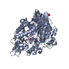
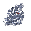
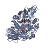
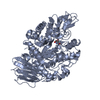
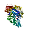
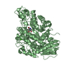
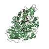
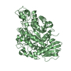
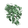
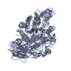
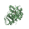
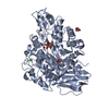
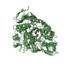
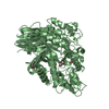
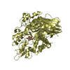
- PDB-4ha1: MutB inactive double mutant D200A-D415N in complex with isomaltulose -
+ Open data

- Basic information
| Entry | Database: PDB / ID: 4ha1 | |||||||||
|---|---|---|---|---|---|---|---|---|---|---|
| Title | MutB inactive double mutant D200A-D415N in complex with isomaltulose | |||||||||
 Components Components | Sucrose isomerase | |||||||||
 Keywords Keywords |  ISOMERASE / ISOMALTULOSE SYNTHASE LIKE / TIM-BARREL / (BETA/ALPHA)8 / SUCROSE ISOMERASE / GLYCOSIDE HYDROLASE / TREHALULOSE SYNTHASE / GH13 FAMILY (CAZY DATABASE) / CALCIUM BINDING ISOMERASE / ISOMALTULOSE SYNTHASE LIKE / TIM-BARREL / (BETA/ALPHA)8 / SUCROSE ISOMERASE / GLYCOSIDE HYDROLASE / TREHALULOSE SYNTHASE / GH13 FAMILY (CAZY DATABASE) / CALCIUM BINDING | |||||||||
| Function / homology |  Function and homology information Function and homology information | |||||||||
| Biological species |  Rhizobium (bacteria) Rhizobium (bacteria) | |||||||||
| Method | X-RAY DIFFRACTION / SYNCHROTRON / FOURIER SYNTHESIS / Resolution: 2.2 Å | |||||||||
 Authors Authors | Lipski, A. / Haser, R. / Aghajari, N. | |||||||||
 Citation Citation | Journal: To be Published Title: Insights into product binding in sucrose isomerases from crystal structures of MutB from Rhizobium sp. Authors: Lipski, A. / Watzlawick, H. / Ravaud, S. / Robert, X. / Haser, R. / Mattes, R. / Aghajari, N. | |||||||||
| History |
|
- Structure visualization
| Structure viewer | Molecule: MolmilJmol/JSmol |
|---|
- Downloads & links
-Download
| PDBx/mmCIF format | 4ha1.cif.gz | 263.1 KB | Display | PDBx/mmCIF format |
|---|---|---|---|---|
| PDB format | pdb4ha1.ent.gz | 204.7 KB | Display | PDB format |
| PDBx/mmJSON format | 4ha1.json.gz | Tree view | PDBx/mmJSON format | |
| Others |  Other downloads Other downloads |
-Validation report
| Arichive directory | https://data.pdbj.org/pub/pdb/validation_reports/ha/4ha1ftp://data.pdbj.org/pub/pdb/validation_reports/ha/4ha1 | HTTPS FTP |
|---|
-Related structure data
| Related structure data | 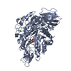 4h7vC 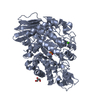 4h8hC 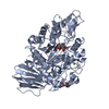 4h8uC 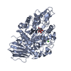 4h8vC 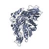 2pwhS S: Starting model for refinement C: citing same article ( |
|---|---|
| Similar structure data |


-Links
 PDBj
PDBj


- Assembly
| Deposited unit | 
| ||||||||
|---|---|---|---|---|---|---|---|---|---|
| 1 | 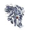
| ||||||||
| 2 | 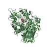
| ||||||||
| Unit cell |
|
-Components
| #1: Protein | Mass: 63893.855 Da / Num. of mol.: 2 / Fragment: TREHALULOSE SYNTHASE MUTB, UNP residues 28-584 / Mutation: D200A, D415N Source method: isolated from a genetically manipulated source Source: (gene. exp.) Rhizobium (bacteria) / Strain: MX-45 / Gene: mutB / Plasmid: pHWG800.2 / Production host: Escherichia coli (E. coli) / Strain (production host): JM109References: UniProt: Q2PS28, UniProt: M1E1F6*PLUS, EC: 5.4.11.99 #2: Chemical |   Mass: 40.078 Da / Num. of mol.: 2 / Source method: obtained synthetically / Formula: Ca Mass: 40.078 Da / Num. of mol.: 2 / Source method: obtained synthetically / Formula: Ca#3: Sugar | ChemComp-ISL / | Isomaltulose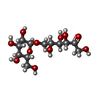  Type: D-saccharide / Mass: 342.296 Da / Num. of mol.: 1 Type: D-saccharide / Mass: 342.296 Da / Num. of mol.: 1Source method: isolated from a genetically manipulated source Formula: C12H22O11 #4: Sugar | ChemComp-GLC / | Glucose  Type: D-saccharide, alpha linking / Mass: 180.156 Da / Num. of mol.: 1 Type: D-saccharide, alpha linking / Mass: 180.156 Da / Num. of mol.: 1Source method: isolated from a genetically manipulated source Formula: C6H12O6 #5: Water | ChemComp-HOH / | Water Mass: 18.015 Da / Num. of mol.: 1162 / Source method: isolated from a natural source / Formula: H2O Mass: 18.015 Da / Num. of mol.: 1162 / Source method: isolated from a natural source / Formula: H2O |
|---|
-Experimental details
-Experiment
| Experiment | Method: X-RAY DIFFRACTION / Number of used crystals: 1 |
|---|
- Sample preparation
| Crystal | Density Matthews: 2.63 Å3/Da / Density % sol: 53.22 % |
|---|---|
| Crystal grow | Temperature: 292 K / Method: vapor diffusion, hanging drop / pH: 8.5 Details: 20% PEG10000, 0.1M TRIS-HCL , pH 8.5, VAPOR DIFFUSION, HANGING DROP, temperature 292K |
-Data collection
| Diffraction | Mean temperature: 100 K |
|---|---|
| Diffraction source | Source: SYNCHROTRON / Site: ESRF  / Beamline: ID23-2 / Wavelength: 0.8726 Å / Beamline: ID23-2 / Wavelength: 0.8726 Å |
| Detector | Type: MARMOSAIC 225 mm CCD / Detector: CCD / Date: Sep 26, 2009 |
| Radiation | Monochromator: Si 111 CHANNEL / Protocol: SINGLE WAVELENGTH / Monochromatic (M) / Laue (L): M / Scattering type: x-ray |
| Radiation wavelength | Wavelength: 0.8726 Å / Relative weight: 1 |
| Reflection | Resolution: 2.2→43.4 Å / Num. all: 66205 / Num. obs: 64580 / % possible obs: 97.5 % / Observed criterion σ(F): 0 / Observed criterion σ(I): 1 / Redundancy: 3.1 % / Biso Wilson estimate: 24.39 Å2 / Rsym value: 0.165 / Net I/σ(I): 8.49 |
| Reflection shell | Resolution: 2.2→2.5 Å / Redundancy: 3.2 % / Rmerge(I) obs: 0.441 / Mean I/σ(I) obs: 3.95 / % possible all: 97.1 |
- Processing
| Software |
| ||||||||||||||||||||||||||||||||||||||||||||||||||||||||||||||||||||||||||||||||||||||||||||||||||||||||||||||||||||||||||||||||||||||||||||||||||||||||||||||||||||||||
|---|---|---|---|---|---|---|---|---|---|---|---|---|---|---|---|---|---|---|---|---|---|---|---|---|---|---|---|---|---|---|---|---|---|---|---|---|---|---|---|---|---|---|---|---|---|---|---|---|---|---|---|---|---|---|---|---|---|---|---|---|---|---|---|---|---|---|---|---|---|---|---|---|---|---|---|---|---|---|---|---|---|---|---|---|---|---|---|---|---|---|---|---|---|---|---|---|---|---|---|---|---|---|---|---|---|---|---|---|---|---|---|---|---|---|---|---|---|---|---|---|---|---|---|---|---|---|---|---|---|---|---|---|---|---|---|---|---|---|---|---|---|---|---|---|---|---|---|---|---|---|---|---|---|---|---|---|---|---|---|---|---|---|---|---|---|---|---|---|---|
| Refinement | Method to determine structure: FOURIER SYNTHESIS Starting model: 2PWH Resolution: 2.2→43.36 Å / Cor.coef. Fo:Fc: 0.939 / Cor.coef. Fo:Fc free: 0.873 / SU ML: 0.28 / σ(F): 1.99 / Phase error: 27.4 / Stereochemistry target values: ML
| ||||||||||||||||||||||||||||||||||||||||||||||||||||||||||||||||||||||||||||||||||||||||||||||||||||||||||||||||||||||||||||||||||||||||||||||||||||||||||||||||||||||||
| Solvent computation | Shrinkage radii: 0.9 Å / VDW probe radii: 1.11 Å / Solvent model: FLAT BULK SOLVENT MODEL | ||||||||||||||||||||||||||||||||||||||||||||||||||||||||||||||||||||||||||||||||||||||||||||||||||||||||||||||||||||||||||||||||||||||||||||||||||||||||||||||||||||||||
| Displacement parameters | Biso mean: 18.44 Å2
| ||||||||||||||||||||||||||||||||||||||||||||||||||||||||||||||||||||||||||||||||||||||||||||||||||||||||||||||||||||||||||||||||||||||||||||||||||||||||||||||||||||||||
| Refinement step | Cycle: LAST / Resolution: 2.2→43.36 Å
| ||||||||||||||||||||||||||||||||||||||||||||||||||||||||||||||||||||||||||||||||||||||||||||||||||||||||||||||||||||||||||||||||||||||||||||||||||||||||||||||||||||||||
| Refine LS restraints |
| ||||||||||||||||||||||||||||||||||||||||||||||||||||||||||||||||||||||||||||||||||||||||||||||||||||||||||||||||||||||||||||||||||||||||||||||||||||||||||||||||||||||||
| LS refinement shell |
|
