 Movie
Movie Controller

[English] 日本語
 Yorodumi
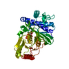
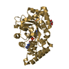
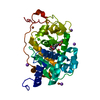
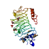
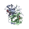
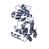
- PDB-4h0a: Crystal structure of a cysteine-rich secretory protein (SAV1118) ... -
+ Open data

- Basic information
| Entry | Database: PDB / ID: 4h0a | ||||||
|---|---|---|---|---|---|---|---|
| Title | Crystal structure of a cysteine-rich secretory protein (SAV1118) from Staphylococcus aureus subsp. aureus Mu50 at 1.90 A resolution | ||||||
 Components Components | Uncharacterized protein | ||||||
 Keywords Keywords | UNKNOWN FUNCTION / CAP protein family / cysteine-rich secretory proteins /  Structural Genomics / Joint Center for Structural Genomics / JCSG / Protein Structure Initiative / PSI-BIOLOGY Structural Genomics / Joint Center for Structural Genomics / JCSG / Protein Structure Initiative / PSI-BIOLOGY | ||||||
| Function / homology |  Function and homology information Function and homology information | ||||||
| Biological species |   Staphylococcus aureus subsp. aureus (bacteria) Staphylococcus aureus subsp. aureus (bacteria) | ||||||
| Method | X-RAY DIFFRACTION / SYNCHROTRON / MAD / Resolution: 1.9 Å | ||||||
 Authors Authors | Joint Center for Structural Genomics (JCSG) | ||||||
 Citation Citation | Journal: To be published Title: Crystal structure of a hypothetical protein (SAV1118) from Staphylococcus aureus subsp. aureus Mu50 at 1.90 A resolution Authors: Joint Center for Structural Genomics (JCSG) | ||||||
| History |
|
- Structure visualization
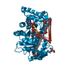
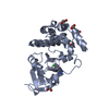
| Structure viewer | Molecule: MolmilJmol/JSmol |
|---|
- Downloads & links
-Download
| PDBx/mmCIF format | 4h0a.cif.gz | 260.7 KB | Display | PDBx/mmCIF format |
|---|---|---|---|---|
| PDB format | pdb4h0a.ent.gz | 215.2 KB | Display | PDB format |
| PDBx/mmJSON format | 4h0a.json.gz | Tree view | PDBx/mmJSON format | |
| Others |  Other downloads Other downloads |
-Validation report
| Arichive directory | https://data.pdbj.org/pub/pdb/validation_reports/h0/4h0aftp://data.pdbj.org/pub/pdb/validation_reports/h0/4h0a | HTTPS FTP |
|---|
-Related structure data
| Similar structure data | |
|---|---|
| Other databases |

-Links
 PDBj
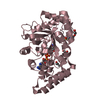
PDBj- Assembly
| Deposited unit | 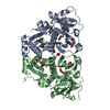
| ||||||||
|---|---|---|---|---|---|---|---|---|---|
| 1 | 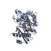
| ||||||||
| 2 | 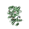
| ||||||||
| Unit cell |
| ||||||||
| Details | CRYSTAL PACKING ANALYSIS SUGGESTS THE ASSIGNMENT OF A MONOMER AS THE SIGNIFICANT OLIGOMERIZATION STATE. |
-Components
| #1: Protein | Mass: 37228.766 Da / Num. of mol.: 2 / Fragment: UNP residues 24-345 Source method: isolated from a genetically manipulated source Source: (gene. exp.) Staphylococcus aureus subsp. aureus (bacteria)Strain: Mu50 / ATCC 700699 / Gene: SAV1118 / Plasmid: SpeedET / Production host: Escherichia coli (E. coli) / Strain (production host): PB1 / References: UniProt: Q99UY5, UniProt: A0A0H3JR67*PLUS#2: Chemical | ChemComp-EDO / Ethylene glycol  Mass: 62.068 Da / Num. of mol.: 5 / Source method: obtained synthetically / Formula: C2H6O2 Mass: 62.068 Da / Num. of mol.: 5 / Source method: obtained synthetically / Formula: C2H6O2#3: Water | ChemComp-HOH / | Water Mass: 18.015 Da / Num. of mol.: 276 / Source method: isolated from a natural source / Formula: H2O Mass: 18.015 Da / Num. of mol.: 276 / Source method: isolated from a natural source / Formula: H2OSequence details | THIS CONSTRUCT WAS EXPRESSED WITH A PURIFICATION TAG MGSDKIHHHHHHENLYFQG. THE TAG WAS REMOVED WITH ...THIS CONSTRUCT WAS EXPRESSED WITH A PURIFICATI | |
|---|
-Experimental details
-Experiment
| Experiment | Method: X-RAY DIFFRACTION / Number of used crystals: 1 |
|---|
- Sample preparation
| Crystal | Density Matthews: 2.28 Å3/Da / Density % sol: 46 % |
|---|---|
| Crystal grow | Temperature: 277 K / Method: vapor diffusion, sitting drop / pH: 4 Details: 1.00M LiCl, 10.00% PEG-6000, 0.1M Citrate pH 4.0, NANODROP, VAPOR DIFFUSION, SITTING DROP, temperature 277K |
-Data collection
| Diffraction | Mean temperature: 100 K | |||||||||||||||||||||||||||||||||||||||||||||||||||||||||||||||||||||||||||||
|---|---|---|---|---|---|---|---|---|---|---|---|---|---|---|---|---|---|---|---|---|---|---|---|---|---|---|---|---|---|---|---|---|---|---|---|---|---|---|---|---|---|---|---|---|---|---|---|---|---|---|---|---|---|---|---|---|---|---|---|---|---|---|---|---|---|---|---|---|---|---|---|---|---|---|---|---|---|---|
| Diffraction source | Source: SYNCHROTRON / Site: ALS  / Beamline: 8.2.2 / Wavelength: 0.9464,0.9795,0.9793 / Beamline: 8.2.2 / Wavelength: 0.9464,0.9795,0.9793 | |||||||||||||||||||||||||||||||||||||||||||||||||||||||||||||||||||||||||||||
| Detector | Type: ADSC QUANTUM 315 / Detector: CCD / Date: Oct 15, 2011 / Details: KOHZU: Double Crystal Si(111) | |||||||||||||||||||||||||||||||||||||||||||||||||||||||||||||||||||||||||||||
| Radiation | Monochromator: Double Crystal Si(111) / Protocol: MAD / Monochromatic (M) / Laue (L): M / Scattering type: x-ray | |||||||||||||||||||||||||||||||||||||||||||||||||||||||||||||||||||||||||||||
| Radiation wavelength |
| |||||||||||||||||||||||||||||||||||||||||||||||||||||||||||||||||||||||||||||
| Reflection | Resolution: 1.9→29.181 Å / Num. obs: 52080 / % possible obs: 96.3 % / Observed criterion σ(I): -3 / Biso Wilson estimate: 34.616 Å2 / Rmerge(I) obs: 0.036 / Net I/σ(I): 11.38 | |||||||||||||||||||||||||||||||||||||||||||||||||||||||||||||||||||||||||||||
| Reflection shell |
|
-Phasing
| Phasing | Method: MAD |
|---|
- Processing
| Software |
| ||||||||||||||||||||||||||||||||||||||||||||||||||||||||||||||||||||||||||||||||||||||||||||||||||||||||||||
|---|---|---|---|---|---|---|---|---|---|---|---|---|---|---|---|---|---|---|---|---|---|---|---|---|---|---|---|---|---|---|---|---|---|---|---|---|---|---|---|---|---|---|---|---|---|---|---|---|---|---|---|---|---|---|---|---|---|---|---|---|---|---|---|---|---|---|---|---|---|---|---|---|---|---|---|---|---|---|---|---|---|---|---|---|---|---|---|---|---|---|---|---|---|---|---|---|---|---|---|---|---|---|---|---|---|---|---|---|---|
| Refinement | Method to determine structure: MAD / Resolution: 1.9→29.181 Å / Cor.coef. Fo:Fc: 0.9542 / Cor.coef. Fo:Fc free: 0.9374 / Occupancy max: 1 / Occupancy min: 0.4 / Cross valid method: THROUGHOUT / σ(F): 0 Details: 1. ZERO OCCUPANCY HYDROGENS WERE INCLUDED DURING REFINEMENT TO IMPROVE THE ANTI-BUMPING RESTRAINTS. 2. ATOM RECORD CONTAINS SUM OF TLS AND RESIDUAL B FACTORS. 3. ANISOU RECORD CONTAINS SUM ...Details: 1. ZERO OCCUPANCY HYDROGENS WERE INCLUDED DURING REFINEMENT TO IMPROVE THE ANTI-BUMPING RESTRAINTS. 2. ATOM RECORD CONTAINS SUM OF TLS AND RESIDUAL B FACTORS. 3. ANISOU RECORD CONTAINS SUM OF TLS AND RESIDUAL U FACTORS. 4. A MET-INHIBITION PROTOCOL WAS USED FOR SELENOMETHIONINE INCORPORATION DURING PROTEIN EXPRESSION. THE OCCUPANCY OF THE SE ATOMS IN THE MSE RESIDUES WAS REDUCED TO 0.75 FOR THE REDUCED SCATTERING POWER DUE TO PARTIAL S-MET INCORPORATION. 5. NCS RESTRAINTS WERE APPLIED DURING REFINEMENT USING LSSR (-AUTONCS) IN BUSTER. 6. MAD PHASE RESTRAINTS WERE USED DURING REFINEMENT. 7. 1,2-ETHANEDIOL (EDO) MOLECULES FROM THE CRYOPROTECTION SOLUTION ARE MODELED. 8. THERE IS SOME UNMODELED ELECTRON DENSITY IN THE REGION NEAR PHE-87, VAL-304 AND GLN-227 IN BOTH CHAINS. 9. THE N-TERMINAL REGIONS (A29-A34 AND B25-B37) HAVE POOR ELECTRON DENSITY AND MAY CONTAIN REGISTER ERRORS.
| ||||||||||||||||||||||||||||||||||||||||||||||||||||||||||||||||||||||||||||||||||||||||||||||||||||||||||||
| Displacement parameters | Biso max: 190.77 Å2 / Biso mean: 59.4636 Å2 / Biso min: 23.51 Å2
| ||||||||||||||||||||||||||||||||||||||||||||||||||||||||||||||||||||||||||||||||||||||||||||||||||||||||||||
| Refine analyze | Luzzati coordinate error obs: 0.34 Å | ||||||||||||||||||||||||||||||||||||||||||||||||||||||||||||||||||||||||||||||||||||||||||||||||||||||||||||
| Refinement step | Cycle: LAST / Resolution: 1.9→29.181 Å
| ||||||||||||||||||||||||||||||||||||||||||||||||||||||||||||||||||||||||||||||||||||||||||||||||||||||||||||
| Refine LS restraints |
| ||||||||||||||||||||||||||||||||||||||||||||||||||||||||||||||||||||||||||||||||||||||||||||||||||||||||||||
| LS refinement shell | Resolution: 1.9→1.95 Å / Total num. of bins used: 20
| ||||||||||||||||||||||||||||||||||||||||||||||||||||||||||||||||||||||||||||||||||||||||||||||||||||||||||||
| Refinement TLS params. | Method: refined / Refine-ID: X-RAY DIFFRACTION
| ||||||||||||||||||||||||||||||||||||||||||||||||||||||||||||||||||||||||||||||||||||||||||||||||||||||||||||
| Refinement TLS group |
|
