Mass: 709.661 Da / Num. of mol.: 1 / Source method: obtained synthetically
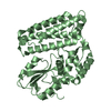
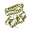
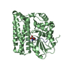
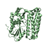
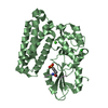
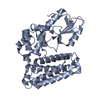
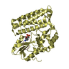
#2: Protein
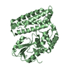
E3ubiquitin-proteinligaseCBL / Casitas B-lineage lymphoma proto-oncogene / Proto-oncogene c-Cbl / RING finger protein 55 / Signal ...Casitas B-lineage lymphoma proto-oncogene / Proto-oncogene c-Cbl / RING finger protein 55 / Signal transduction protein CBL
Mass: 35843.434 Da / Num. of mol.: 1 / Fragment: human Cbl TKB domain residues 47-351 Source method: isolated from a genetically manipulated source Source: (gene. exp.) Homo sapiens (human) / Gene: CBL, CBL2, RNF55 / Plasmid: pET28 / Production host: Escherichia coli (E. coli) / Strain (production host): ROSETTA-2 References: UniProt: P22681, Ligases; Forming carbon-nitrogen bonds; Acid-amino-acid ligases (peptide synthases)
Resolution: 3→32.66 Å / Cor.coef. Fo:Fc: 0.941 / Cor.coef. Fo:Fc free: 0.891 / SU B: 17.092 / SU ML: 0.301 / Cross valid method: THROUGHOUT / ESU R Free: 0.404 / Stereochemistry target values: MAXIMUM LIKELIHOOD / Details: HYDROGENS HAVE BEEN USED IF PRESENT IN THE INPUT
Rfactor
Num. reflection
% reflection
Selection details
Rfree
0.24828
470
4.9 %
RANDOM
Rwork
0.18216
-
-
-
obs
0.18549
9213
98.3 %
-
Solvent computation
Ion probe radii: 0.8 Å / Shrinkage radii: 0.8 Å / VDW probe radii: 1.2 Å / Solvent model: MASK
Displacement parameters
Biso mean: 46.745 Å2
Baniso -1
Baniso -2
Baniso -3
1-
-2 Å2
-1 Å2
0 Å2
2-
-
-2 Å2
0 Å2
3-
-
-
3 Å2
Refinement step
Cycle: LAST / Resolution: 3→32.66 Å
Protein
Nucleic acid
Ligand
Solvent
Total
Num. atoms
2547
0
0
41
2588
Refine LS restraints
Refine-ID
Type
Dev ideal
Dev ideal target
Number
X-RAY DIFFRACTION
r_bond_refined_d
0.012
0.02
2616
X-RAY DIFFRACTION
r_bond_other_d
X-RAY DIFFRACTION
r_angle_refined_deg
1.685
1.971
3537
X-RAY DIFFRACTION
r_angle_other_deg
X-RAY DIFFRACTION
r_dihedral_angle_1_deg
6.497
5
308
X-RAY DIFFRACTION
r_dihedral_angle_2_deg
36.62
23.417
120
X-RAY DIFFRACTION
r_dihedral_angle_3_deg
22.866
15
464
X-RAY DIFFRACTION
r_dihedral_angle_4_deg
19.296
15
17
X-RAY DIFFRACTION
r_chiral_restr
0.106
0.2
375
X-RAY DIFFRACTION
r_gen_planes_refined
0.007
0.021
1974
X-RAY DIFFRACTION
r_gen_planes_other
X-RAY DIFFRACTION
r_nbd_refined
X-RAY DIFFRACTION
r_nbd_other
X-RAY DIFFRACTION
r_nbtor_refined
X-RAY DIFFRACTION
r_nbtor_other
X-RAY DIFFRACTION
r_xyhbond_nbd_refined
X-RAY DIFFRACTION
r_xyhbond_nbd_other
X-RAY DIFFRACTION
r_metal_ion_refined
X-RAY DIFFRACTION
r_metal_ion_other
X-RAY DIFFRACTION
r_symmetry_vdw_refined
X-RAY DIFFRACTION
r_symmetry_vdw_other
X-RAY DIFFRACTION
r_symmetry_hbond_refined
X-RAY DIFFRACTION
r_symmetry_hbond_other
X-RAY DIFFRACTION
r_symmetry_metal_ion_refined
X-RAY DIFFRACTION
r_symmetry_metal_ion_other
X-RAY DIFFRACTION
r_mcbond_it
X-RAY DIFFRACTION
r_mcbond_other
X-RAY DIFFRACTION
r_mcangle_it
X-RAY DIFFRACTION
r_scbond_it
X-RAY DIFFRACTION
r_scangle_it
X-RAY DIFFRACTION
r_rigid_bond_restr
X-RAY DIFFRACTION
r_sphericity_free
X-RAY DIFFRACTION
r_sphericity_bonded
LS refinement shell
Resolution: 3→3.078 Å / Total num. of bins used: 20
Rfactor
Num. reflection
% reflection
Rfree
0.387
30
-
Rwork
0.258
646
-
obs
-
-
100 %
+
About Yorodumi
-
News
-
Feb 9, 2022. New format data for meta-information of EMDB entries
New format data for meta-information of EMDB entries
Version 3 of the EMDB header file is now the official format.
The previous official version 1.9 will be removed from the archive.
In the structure databanks used in Yorodumi, some data are registered as the other names, "COVID-19 virus" and "2019-nCoV". Here are the details of the virus and the list of structure data.
Jan 31, 2019. EMDB accession codes are about to change! (news from PDBe EMDB page)
EMDB accession codes are about to change! (news from PDBe EMDB page)
The allocation of 4 digits for EMDB accession codes will soon come to an end. Whilst these codes will remain in use, new EMDB accession codes will include an additional digit and will expand incrementally as the available range of codes is exhausted. The current 4-digit format prefixed with “EMD-” (i.e. EMD-XXXX) will advance to a 5-digit format (i.e. EMD-XXXXX), and so on. It is currently estimated that the 4-digit codes will be depleted around Spring 2019, at which point the 5-digit format will come into force.
The EM Navigator/Yorodumi systems omit the EMD- prefix.
Related info.:Q: What is EMD? / ID/Accession-code notation in Yorodumi/EM Navigator
Yorodumi is a browser for structure data from EMDB, PDB, SASBDB, etc.
This page is also the successor to EM Navigator detail page, and also detail information page/front-end page for Omokage search.
The word "yorodu" (or yorozu) is an old Japanese word meaning "ten thousand". "mi" (miru) is to see.
Related info.:EMDB / PDB / SASBDB / Comparison of 3 databanks / Yorodumi Search / Aug 31, 2016. New EM Navigator & Yorodumi / Yorodumi Papers / Jmol/JSmol / Function and homology information / Changes in new EM Navigator and Yorodumi
 Movie
Movie Controller


 Open data
 Basic information
 Components
 Keywords
 Function and homology information
 flotillin complex / phosphatidylinositol 3-kinase regulatory subunit binding / positive regulation of epidermal growth factor receptor signaling pathway / Regulation of KIT signaling / mast cell degranulation / Interleukin-6 signaling ...regulation of platelet-derived growth factor receptor-alpha signaling pathway / ubiquitin-dependent endocytosis / regulation of Rap protein signal transduction / entry of bacterium into host cell /
 Authors
 Citation
 Structure visualization
 Downloads & links
 Other downloads
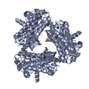

 PDBj
PDBj


 Assembly
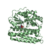

 Mass: 18.015 Da / Num. of mol.: 41 / Source method: isolated from a natural source / Formula: H2O
 Sample preparation
 Processing