 Movie
Movie Controller


+ Open data

- Basic information
| Entry | Database: PDB / ID: 4glc | ||||||||||||||||||||
|---|---|---|---|---|---|---|---|---|---|---|---|---|---|---|---|---|---|---|---|---|---|
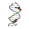
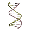
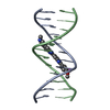
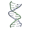
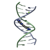
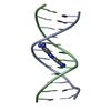
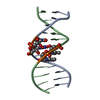
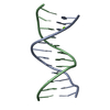
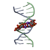
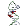
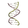
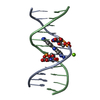
| Title | DNA dodecamer containing 5-hydroxymethyl-cytosine | ||||||||||||||||||||
 Components Components | DNA (5'-D(* Keywords Keywords DNA / B-DNA dodecamer / epigenetics / 5-hydroxymethyl cytosine DNA / B-DNA dodecamer / epigenetics / 5-hydroxymethyl cytosineFunction / homology | DNA / DNA (> 10) Function and homology information Function and homology informationBiological species | Synthetic construct (others) | Method | X-RAY DIFFRACTION / SYNCHROTRON / MOLECULAR REPLACEMENT / Resolution: 1.831 Å  Authors AuthorsSpingler, B. / Renciuk, D. / Vorlickova, M. |  CitationJournal: Nucleic Acids Res. / Year: 2013 CitationJournal: Nucleic Acids Res. / Year: 2013Title: Crystal structures of B-DNA dodecamer containing the epigenetic modifications 5-hydroxymethylcytosine or 5-methylcytosine. Authors: Renciuk, D. / Blacque, O. / Vorlickova, M. / Spingler, B. History |
|
- Structure visualization
| Structure viewer | Molecule: MolmilJmol/JSmol |
|---|
- Downloads & links
-Download
| PDBx/mmCIF format | 4glc.cif.gz | 23.4 KB | Display | PDBx/mmCIF format |
|---|---|---|---|---|
| PDB format | pdb4glc.ent.gz | 14.8 KB | Display | PDB format |
| PDBx/mmJSON format | 4glc.json.gz | Tree view | PDBx/mmJSON format | |
| Others |  Other downloads Other downloads |
-Validation report
| Arichive directory | https://data.pdbj.org/pub/pdb/validation_reports/gl/4glcftp://data.pdbj.org/pub/pdb/validation_reports/gl/4glc | HTTPS FTP |
|---|
-Related structure data
| Related structure data |  4gjuC  4glgC  4glhC  4hliC  1dpnS S: Starting model for refinement C: citing same article ( |
|---|---|
| Similar structure data |


-Links
 PDBj
PDBj


- Assembly
| Deposited unit | 
| ||||||||
|---|---|---|---|---|---|---|---|---|---|
| 1 |
| ||||||||
| Unit cell |
|
-Components
| #1: DNA chain | Mass: 3693.418 Da / Num. of mol.: 2 / Source method: obtained synthetically / Source: (synth.) Synthetic construct (others) #2: Water | ChemComp-HOH / | Water Mass: 18.015 Da / Num. of mol.: 40 / Source method: isolated from a natural source / Formula: H2O Mass: 18.015 Da / Num. of mol.: 40 / Source method: isolated from a natural source / Formula: H2O |
|---|
-Experimental details
-Experiment
| Experiment | Method: X-RAY DIFFRACTION / Number of used crystals: 1 |
|---|
- Sample preparation
| Crystal | Density Matthews: 1.98 Å3/Da / Density % sol: 46.04 % |
|---|---|
| Crystal grow | Temperature: 293 K / pH: 7 Details: 0.04M sodium cacodylate buffer, 0.08M sodium chloride, 0.012M spermine tetra hydrochloride, 30% MPD, pH 7.0, VAPOR DIFFUSION, SITTING DROP, temperature 293K |
-Data collection
| Diffraction | Mean temperature: 100 K |
|---|---|
| Diffraction source | Source: SYNCHROTRON / Site: SLS  / Beamline: X06DA / Wavelength: 1.00067 / Beamline: X06DA / Wavelength: 1.00067 |
| Detector | Type: DECTRIS PILATUS 2M / Detector: PIXEL / Date: Jun 9, 2012 |
| Radiation | Monochromator: BARTELS MONOCHROMATOR / Protocol: SINGLE WAVELENGTH / Monochromatic (M) / Laue (L): M / Scattering type: x-ray |
| Radiation wavelength | Wavelength: 1.00067 Å / Relative weight: 1 |
| Reflection | Resolution: 1.831→41.6 Å / Num. obs: 6201 / % possible obs: 97.5 % / Redundancy: 3.68 % / Rmerge(I) obs: 0.034 / Rsym value: 0.021 / Net I/σ(I): 24.06 |
| Reflection shell | Resolution: 1.831→1.94 Å / Redundancy: 3.62 % / Rmerge(I) obs: 0.216 / Mean I/σ(I) obs: 6.65 / % possible all: 96.6 |
- Processing
| Software |
| |||||||||||||||||||||||||||||||||
|---|---|---|---|---|---|---|---|---|---|---|---|---|---|---|---|---|---|---|---|---|---|---|---|---|---|---|---|---|---|---|---|---|---|---|
| Refinement | Method to determine structure: MOLECULAR REPLACEMENT Starting model: PDB ENTRY 1DPN Resolution: 1.831→35.2 Å / Num. parameters: 2123 / Num. restraintsaints: 2406 / Cross valid method: FREE R / σ(F): 0 / Stereochemistry target values: ENGH AND HUBER
| |||||||||||||||||||||||||||||||||
| Refine analyze | Num. disordered residues: 0 / Occupancy sum hydrogen: 0 / Occupancy sum non hydrogen: 528.6 | |||||||||||||||||||||||||||||||||
| Refinement step | Cycle: LAST / Resolution: 1.831→35.2 Å
| |||||||||||||||||||||||||||||||||
| Refine LS restraints |
|
