 Movie
Movie Controller


+ Open data

- Basic information
| Entry | Database: PDB / ID: 4giw | ||||||
|---|---|---|---|---|---|---|---|
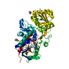
| Title | Crystal structure of the RUN domain of human NESCA | ||||||
 Components Components | RUN and SH3 domain-containing protein 1 | ||||||
 Keywords Keywords |  SIGNALING PROTEIN / NGF-TrkA signaling pathway / NF-kB pathway SIGNALING PROTEIN / NGF-TrkA signaling pathway / NF-kB pathway | ||||||
| Function / homology |  Function and homology information Function and homology informationprotein polyubiquitination / microtubule cytoskeleton / actin binding / cytoplasmic vesicle / microtubule / postsynaptic density / early endosome / Golgi apparatus / nucleus / cytosolSimilarity search - Function | ||||||
| Biological species |  Homo sapiens (human) Homo sapiens (human) | ||||||
| Method | X-RAY DIFFRACTION / SYNCHROTRON / SAD / Resolution: 2 Å | ||||||
 Authors Authors | Bai, L. / Sun, Q. / Jiang, T. | ||||||
 Citation Citation | Journal: Protein Cell / Year: 2012 Title: Crystal structure and functional implication of the RUN domain of human NESCA Authors: Sun, Q. / Han, C. / Liu, L. / Wang, Y. / Deng, H. / Bai, L. / Jiang, T. | ||||||
| History |
|
- Structure visualization
| Structure viewer | Molecule: MolmilJmol/JSmol |
|---|
- Downloads & links
-Download
| PDBx/mmCIF format | 4giw.cif.gz | 76.4 KB | Display | PDBx/mmCIF format |
|---|---|---|---|---|
| PDB format | pdb4giw.ent.gz | 61.8 KB | Display | PDB format |
| PDBx/mmJSON format | 4giw.json.gz | Tree view | PDBx/mmJSON format | |
| Others |  Other downloads Other downloads |
-Validation report
| Arichive directory | https://data.pdbj.org/pub/pdb/validation_reports/gi/4giwftp://data.pdbj.org/pub/pdb/validation_reports/gi/4giw | HTTPS FTP |
|---|
-Related structure data
| Similar structure data |
|---|


-Links
 PDBj
PDBj
- Assembly
| Deposited unit | 
| ||||||||
|---|---|---|---|---|---|---|---|---|---|
| 1 |
| ||||||||
| Unit cell |
|
-Components
| #1: Protein | Mass: 21829.924 Da / Num. of mol.: 2 / Fragment: RUN domain / Mutation: L150M, L152M Source method: isolated from a genetically manipulated source Source: (gene. exp.) Homo sapiens (human) / Gene: NESCA / Production host:  Escherichia coli (E. coli) / References: UniProt: Q9BVN2 Escherichia coli (E. coli) / References: UniProt: Q9BVN2#2: Water | ChemComp-HOH / | Water Mass: 18.015 Da / Num. of mol.: 149 / Source method: isolated from a natural source / Formula: H2O Mass: 18.015 Da / Num. of mol.: 149 / Source method: isolated from a natural source / Formula: H2O |
|---|
-Experimental details
-Experiment
| Experiment | Method: X-RAY DIFFRACTION / Number of used crystals: 1 |
|---|
- Sample preparation
| Crystal | Density Matthews: 1.86 Å3/Da / Density % sol: 33.73 % |
|---|---|
| Crystal grow | Temperature: 289 K / Method: vapor diffusion, sitting drop Details: 20%(w/v) PEG3350, 0.2M K2HPO4, VAPOR DIFFUSION, SITTING DROP, temperature 289K |
-Data collection
| Diffraction | Mean temperature: 100 K |
|---|---|
| Diffraction source | Source: SYNCHROTRON / Site: SSRF  / Beamline: BL17U / Wavelength: 0.97915 Å / Beamline: BL17U / Wavelength: 0.97915 Å |
| Detector | Type: ADSC QUANTUM 315r / Detector: CCD / Date: Jun 10, 2011 |
| Radiation | Monochromator: double crystal / Protocol: SINGLE WAVELENGTH / Monochromatic (M) / Laue (L): M / Scattering type: x-ray |
| Radiation wavelength | Wavelength: 0.97915 Å / Relative weight: 1 |
| Reflection | Resolution: 2→35.7 Å / Num. all: 20352 / Num. obs: 20352 / % possible obs: 99.63 % / Observed criterion σ(F): 0 / Observed criterion σ(I): 0 / Redundancy: 7.247 % / Rmerge(I) obs: 0.052 |
| Reflection shell | Resolution: 2→2.07 Å / Redundancy: 7.2 % / Rmerge(I) obs: 0.103 / Mean I/σ(I) obs: 2.8 / Num. unique all: 2144 / % possible all: 99.42 |
- Processing
| Software |
| |||||||||||||||||||||||||||||||||||||||||||||||||||||||||||||||||
|---|---|---|---|---|---|---|---|---|---|---|---|---|---|---|---|---|---|---|---|---|---|---|---|---|---|---|---|---|---|---|---|---|---|---|---|---|---|---|---|---|---|---|---|---|---|---|---|---|---|---|---|---|---|---|---|---|---|---|---|---|---|---|---|---|---|---|
| Refinement | Method to determine structure: SAD / Resolution: 2→35.7 Å / Cor.coef. Fo:Fc: 0.924 / Cor.coef. Fo:Fc free: 0.921 / SU B: 2.873 / SU ML: 0.084 / Cross valid method: THROUGHOUT / ESU R: 0.234 / ESU R Free: 0.162 / Stereochemistry target values: MAXIMUM LIKELIHOOD Details: HYDROGENS HAVE BEEN ADDED IN THE RIDING POSITIONS. THE STRUCTURE WAS REFINED ALSO WITH PHENIX.
| |||||||||||||||||||||||||||||||||||||||||||||||||||||||||||||||||
| Solvent computation | Ion probe radii: 0.8 Å / Shrinkage radii: 0.8 Å / VDW probe radii: 1.4 Å / Solvent model: MASK | |||||||||||||||||||||||||||||||||||||||||||||||||||||||||||||||||
| Displacement parameters | Biso mean: 16.251 Å2
| |||||||||||||||||||||||||||||||||||||||||||||||||||||||||||||||||
| Refinement step | Cycle: LAST / Resolution: 2→35.7 Å
| |||||||||||||||||||||||||||||||||||||||||||||||||||||||||||||||||
| Refine LS restraints |
| |||||||||||||||||||||||||||||||||||||||||||||||||||||||||||||||||
| LS refinement shell | Resolution: 2.002→2.053 Å / Total num. of bins used: 20
|
