 Movie
Movie Controller


+ Open data

- Basic information
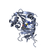
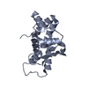
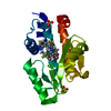
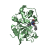
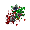
| Entry | Database: PDB / ID: 4geq | ||||||
|---|---|---|---|---|---|---|---|
| Title | Crystal structure of the Spc24-Spc25/Cnn1 binding interface | ||||||
 Components Components |
| ||||||
 Keywords Keywords |  CELL CYCLE / protein-protein complex / Ndc80-binding motif / RWD domain / kinetochore components / nucleus CELL CYCLE / protein-protein complex / Ndc80-binding motif / RWD domain / kinetochore components / nucleus | ||||||
| Function / homology |  Function and homology information Function and homology informationcentromere clustering / negative regulation of kinetochore assembly / Ndc80 complex / sister chromatid biorientation / centromeric DNA binding / chromosome segregation / kinetochore / cell division / protein-containing complex binding / identical protein binding / nucleusSimilarity search - Function | ||||||
| Biological species |  Saccharomyces cerevisiae S288c (yeast) Saccharomyces cerevisiae S288c (yeast) | ||||||
| Method | X-RAY DIFFRACTION / SYNCHROTRON / MOLECULAR REPLACEMENT / Resolution: 2.01 Å | ||||||
 Authors Authors | Malvezzi, F. / Litos, G. / Schleiffer, A. / Heuck, A. / Clausen, T. / Westermann, S. | ||||||
 Citation Citation | Journal: Embo J. / Year: 2013 Title: A structural basis for kinetochore recruitment of the Ndc80 complex via two distinct centromere receptors. Authors: Malvezzi, F. / Litos, G. / Schleiffer, A. / Heuck, A. / Mechtler, K. / Clausen, T. / Westermann, S. | ||||||
| History |
|
- Structure visualization
| Structure viewer | Molecule: MolmilJmol/JSmol |
|---|
- Downloads & links
-Download
| PDBx/mmCIF format | 4geq.cif.gz | 146.3 KB | Display | PDBx/mmCIF format |
|---|---|---|---|---|
| PDB format | pdb4geq.ent.gz | 114.9 KB | Display | PDB format |
| PDBx/mmJSON format | 4geq.json.gz | Tree view | PDBx/mmJSON format | |
| Others |  Other downloads Other downloads |
-Validation report
| Arichive directory | https://data.pdbj.org/pub/pdb/validation_reports/ge/4geqftp://data.pdbj.org/pub/pdb/validation_reports/ge/4geq | HTTPS FTP |
|---|
-Related structure data
| Related structure data |  2ftxS S: Starting model for refinement |
|---|---|
| Similar structure data |


-Links
 PDBj
PDBj- Assembly
| Deposited unit | 
| ||||||||
|---|---|---|---|---|---|---|---|---|---|
| 1 | 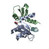
| ||||||||
| 2 | 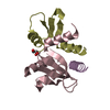
| ||||||||
| Unit cell |
|
-Components
| #1: Protein | Mass: 9889.295 Da / Num. of mol.: 2 / Fragment: Spc25p C-terminal domain, residues 133-221 Source method: isolated from a genetically manipulated source Source: (gene. exp.) Saccharomyces cerevisiae S288c (yeast) / Strain: ATCC 204508 / S288c / Gene: SPC25, YER018C / Plasmid: pETduett-1 / Production host:  Escherichia coli (E. coli) / References: UniProt: P40014 Escherichia coli (E. coli) / References: UniProt: P40014#2: Protein | Mass: 7903.027 Da / Num. of mol.: 2 / Fragment: Spc24p C-terminal domain, residues 155-213 Source method: isolated from a genetically manipulated source Source: (gene. exp.) Saccharomyces cerevisiae S288c (yeast) / Strain: ATCC 204508 / S288c / Gene: SPC24, YM9718.16C, YMR117C / Plasmid: pETduett-1 / Production host: Escherichia coli (E. coli) / References: UniProt: Q04477#3: Protein/peptide | Mass: 2848.133 Da / Num. of mol.: 2 / Fragment: Cnn1p N-terminal motif, residues 60-84 / Source method: obtained synthetically / Source: (synth.) Saccharomyces cerevisiae S288c (yeast) / References: UniProt: P43618#4: Chemical | Glycerol  Mass: 92.094 Da / Num. of mol.: 2 / Source method: obtained synthetically / Formula: C3H8O3 Mass: 92.094 Da / Num. of mol.: 2 / Source method: obtained synthetically / Formula: C3H8O3#5: Water | ChemComp-HOH / | Water Mass: 18.015 Da / Num. of mol.: 150 / Source method: isolated from a natural source / Formula: H2O Mass: 18.015 Da / Num. of mol.: 150 / Source method: isolated from a natural source / Formula: H2O |
|---|
-Experimental details
-Experiment
| Experiment | Method: X-RAY DIFFRACTION / Number of used crystals: 1 |
|---|
- Sample preparation
| Crystal | Density Matthews: 1.78 Å3/Da / Density % sol: 30.83 % |
|---|---|
| Crystal grow | Temperature: 293.15 K / Method: vapor diffusion, sitting drop / pH: 7.6 Details: 15% PEG6000, 5% glycerol; Drop volume: 0.2ul; Protein proportion: 50%; Protein concentration: 6 mg/ml, pH 7.6, VAPOR DIFFUSION, SITTING DROP, temperature 293.15K |
-Data collection
| Diffraction | Mean temperature: 100 K |
|---|---|
| Diffraction source | Source: SYNCHROTRON / Site: PETRA III, EMBL c/o DESY  / Beamline: P13 (MX1) / Wavelength: 1.127 Å / Beamline: P13 (MX1) / Wavelength: 1.127 Å |
| Detector | Type: DECTRIS PILATUS 6M / Detector: PIXEL / Date: Oct 2, 2011 |
| Radiation | Monochromator: Si(111) monochromator / Protocol: SINGLE WAVELENGTH / Monochromatic (M) / Laue (L): M / Scattering type: x-ray |
| Radiation wavelength | Wavelength: 1.127 Å / Relative weight: 1 |
| Reflection | Resolution: 2→61.85 Å / Num. all: 20020 / Num. obs: 19858 / % possible obs: 96.9 % / Observed criterion σ(I): 3 / Redundancy: 4.6 % / Biso Wilson estimate: 24.44 Å2 / Rmerge(I) obs: 0.113 / Net I/σ(I): 8.5 |
| Reflection shell | Resolution: 2→2.11 Å / Redundancy: 3.1 % / Rmerge(I) obs: 0.577 / Mean I/σ(I) obs: 2.5 / % possible all: 87.1 |
- Processing
| Software |
| |||||||||||||||||||||||||||||||||||||||||||||||||||||||||||||||||||||||||||||||||||||||||||||||||||||||||||||||||||||||||||||||||||||||||||||||||||||||||||||||||||||||||||||||
|---|---|---|---|---|---|---|---|---|---|---|---|---|---|---|---|---|---|---|---|---|---|---|---|---|---|---|---|---|---|---|---|---|---|---|---|---|---|---|---|---|---|---|---|---|---|---|---|---|---|---|---|---|---|---|---|---|---|---|---|---|---|---|---|---|---|---|---|---|---|---|---|---|---|---|---|---|---|---|---|---|---|---|---|---|---|---|---|---|---|---|---|---|---|---|---|---|---|---|---|---|---|---|---|---|---|---|---|---|---|---|---|---|---|---|---|---|---|---|---|---|---|---|---|---|---|---|---|---|---|---|---|---|---|---|---|---|---|---|---|---|---|---|---|---|---|---|---|---|---|---|---|---|---|---|---|---|---|---|---|---|---|---|---|---|---|---|---|---|---|---|---|---|---|---|---|---|
| Refinement | Method to determine structure: MOLECULAR REPLACEMENT Starting model: PDB ENTRY 2FTX Resolution: 2.01→37.09 Å / Cor.coef. Fo:Fc: 0.9472 / Cor.coef. Fo:Fc free: 0.9174 / SU R Cruickshank DPI: 0.236 / Cross valid method: THROUGHOUT / σ(F): 0 Stereochemistry target values: Engh & Huber, buster common-compounds v 1.0
| |||||||||||||||||||||||||||||||||||||||||||||||||||||||||||||||||||||||||||||||||||||||||||||||||||||||||||||||||||||||||||||||||||||||||||||||||||||||||||||||||||||||||||||||
| Displacement parameters | Biso mean: 37.25 Å2
| |||||||||||||||||||||||||||||||||||||||||||||||||||||||||||||||||||||||||||||||||||||||||||||||||||||||||||||||||||||||||||||||||||||||||||||||||||||||||||||||||||||||||||||||
| Refine analyze | Luzzati coordinate error obs: 0.325 Å | |||||||||||||||||||||||||||||||||||||||||||||||||||||||||||||||||||||||||||||||||||||||||||||||||||||||||||||||||||||||||||||||||||||||||||||||||||||||||||||||||||||||||||||||
| Refinement step | Cycle: LAST / Resolution: 2.01→37.09 Å
| |||||||||||||||||||||||||||||||||||||||||||||||||||||||||||||||||||||||||||||||||||||||||||||||||||||||||||||||||||||||||||||||||||||||||||||||||||||||||||||||||||||||||||||||
| Refine LS restraints |
| |||||||||||||||||||||||||||||||||||||||||||||||||||||||||||||||||||||||||||||||||||||||||||||||||||||||||||||||||||||||||||||||||||||||||||||||||||||||||||||||||||||||||||||||
| LS refinement shell | Resolution: 2.01→2.12 Å / Total num. of bins used: 10
| |||||||||||||||||||||||||||||||||||||||||||||||||||||||||||||||||||||||||||||||||||||||||||||||||||||||||||||||||||||||||||||||||||||||||||||||||||||||||||||||||||||||||||||||
| Refinement TLS params. | Method: refined / Refine-ID: X-RAY DIFFRACTION
| |||||||||||||||||||||||||||||||||||||||||||||||||||||||||||||||||||||||||||||||||||||||||||||||||||||||||||||||||||||||||||||||||||||||||||||||||||||||||||||||||||||||||||||||
| Refinement TLS group |
|
