 Movie
Movie Controller


+ Open data

- Basic information
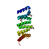
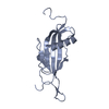
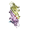
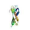
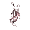
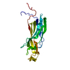
| Entry | Database: PDB / ID: 4ga0 | ||||||
|---|---|---|---|---|---|---|---|
| Title | Structure of the N-terminal domain of Nup358 | ||||||
 Components Components | E3 SUMO-protein ligase RanBP2 | ||||||
 Keywords Keywords |  TRANSPORT PROTEIN / TPR motif / Nuclear pore complex component Nucleocytoplasmic transport TRANSPORT PROTEIN / TPR motif / Nuclear pore complex component Nucleocytoplasmic transport | ||||||
| Function / homology |  Function and homology information Function and homology informationcytoplasmic periphery of the nuclear pore complex / SUMO ligase activity / SUMO ligase complex / annulate lamellae / nuclear pore cytoplasmic filaments / Nuclear Pore Complex (NPC) Disassembly / nuclear inclusion body / nuclear pore nuclear basket / Transport of Ribonucleoproteins into the Host Nucleus / Regulation of Glucokinase by Glucokinase Regulatory Protein ...cytoplasmic periphery of the nuclear pore complex / SUMO ligase activity / SUMO ligase complex / annulate lamellae / nuclear pore cytoplasmic filaments / Nuclear Pore Complex (NPC) Disassembly / nuclear inclusion body / nuclear pore nuclear basket / Transport of Ribonucleoproteins into the Host Nucleus / Regulation of Glucokinase by Glucokinase Regulatory Protein / Defective TPR may confer susceptibility towards thyroid papillary carcinoma (TPC) / Transport of the SLBP independent Mature mRNA / Transport of the SLBP Dependant Mature mRNA / NS1 Mediated Effects on Host Pathways / SUMOylation of SUMOylation proteins / Transport of Mature mRNA Derived from an Intronless Transcript / Transferases; Acyltransferases; Aminoacyltransferases / Rev-mediated nuclear export of HIV RNA / SUMOylation of RNA binding proteins / nuclear export / Nuclear import of Rev protein / Transport of Mature mRNA derived from an Intron-Containing Transcript / NEP/NS2 Interacts with the Cellular Export Machinery / tRNA processing in the nucleus / SUMO transferase activity / nucleocytoplasmic transport / centrosome localization / Viral Messenger RNA Synthesis / regulation of gluconeogenesis / NLS-bearing protein import into nucleus / SUMOylation of ubiquitinylation proteins / Vpr-mediated nuclear import of PICs / SUMOylation of DNA replication proteins / protein sumoylation / Regulation of HSF1-mediated heat shock response / mRNA transport / Amplification of signal from unattached kinetochores via a MAD2 inhibitory signal / SUMOylation of DNA damage response and repair proteins / nuclear pore / Mitotic Prometaphase / EML4 and NUDC in mitotic spindle formation / Resolution of Sister Chromatid Cohesion / response to amphetamine / SUMOylation of chromatin organization proteins / GTPase activator activity / HCMV Late Events / RHO GTPases Activate Formins / Transcriptional regulation by small RNAs / Signaling by ALK fusions and activated point mutants / ISG15 antiviral mechanism / small GTPase binding / HCMV Early Events / Separation of Sister Chromatids / protein folding / nuclear envelope / snRNP Assembly / nuclear membrane / intracellular membrane-bounded organelle / protein-containing complex binding / SARS-CoV-2 activates/modulates innate and adaptive immune responses / RNA binding / nucleoplasm / membrane / metal ion binding / cytosol / cytoplasmSimilarity search - Function | ||||||
| Biological species |  Homo sapiens (human) Homo sapiens (human) | ||||||
| Method | X-RAY DIFFRACTION / SYNCHROTRON / MOLECULAR REPLACEMENT / molecular replacement / Resolution: 1.15 Å | ||||||
 Authors Authors | Kassube, S.A. / Lin, D.H. / Stuwe, T. / Hoelz, A. | ||||||
 Citation Citation | Journal: J.Mol.Biol. / Year: 2012 Title: Crystal structure of the N-terminal domain of Nup358/RanBP2. Authors: Kassube, S.A. / Stuwe, T. / Lin, D.H. / Antonuk, C.D. / Napetschnig, J. / Blobel, G. / Hoelz, A. | ||||||
| History |
|
- Structure visualization
| Structure viewer | Molecule: MolmilJmol/JSmol |
|---|
- Downloads & links
-Download
| PDBx/mmCIF format | 4ga0.cif.gz | 110.8 KB | Display | PDBx/mmCIF format |
|---|---|---|---|---|
| PDB format | pdb4ga0.ent.gz | 88.8 KB | Display | PDB format |
| PDBx/mmJSON format | 4ga0.json.gz | Tree view | PDBx/mmJSON format | |
| Others |  Other downloads Other downloads |
-Validation report
| Arichive directory | https://data.pdbj.org/pub/pdb/validation_reports/ga/4ga0ftp://data.pdbj.org/pub/pdb/validation_reports/ga/4ga0 | HTTPS FTP |
|---|
-Related structure data
| Related structure data |  4ga1C 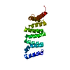 4ga2SC C: citing same article ( S: Starting model for refinement |
|---|---|
| Similar structure data |


-Links
 PDBj
PDBj


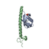
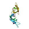
- Assembly
| Deposited unit | 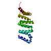
| ||||||||
|---|---|---|---|---|---|---|---|---|---|
| 1 |
| ||||||||
| Unit cell |
|
-Components
| #1: Protein | Mass: 17354.885 Da / Num. of mol.: 1 / Fragment: unp residues 1-145 Source method: isolated from a genetically manipulated source Source: (gene. exp.) Homo sapiens (human) / Gene: RANBP2, NUP358 / Production host:  Escherichia coli (E. coli) / Strain (production host): BL21(DE3) RIL / References: UniProt: P49792 Escherichia coli (E. coli) / Strain (production host): BL21(DE3) RIL / References: UniProt: P49792 |
|---|---|
| #2: Water | ChemComp-HOH / Water Mass: 18.015 Da / Num. of mol.: 85 / Source method: isolated from a natural source / Formula: H2O Mass: 18.015 Da / Num. of mol.: 85 / Source method: isolated from a natural source / Formula: H2O |
-Experimental details
-Experiment
| Experiment | Method: X-RAY DIFFRACTION / Number of used crystals: 1 |
|---|
- Sample preparation
| Crystal | Density Matthews: 2.09 Å3/Da / Density % sol: 41.03 % |
|---|---|
| Crystal grow | Temperature: 277 K / Method: vapor diffusion, hanging drop Details: 18 % (w/v) PEG 3350 200 mM lithium acetate, VAPOR DIFFUSION, HANGING DROP, temperature 277K |
-Data collection
| Diffraction | Mean temperature: 100 K |
|---|---|
| Diffraction source | Source: SYNCHROTRON / Site: SSRL  / Beamline: BL12-2 / Wavelength: 1.0332 Å / Beamline: BL12-2 / Wavelength: 1.0332 Å |
| Detector | Type: DECTRIS PILATUS 6M / Detector: PIXEL / Date: Jan 6, 2012 |
| Radiation | Protocol: SINGLE WAVELENGTH / Monochromatic (M) / Laue (L): M / Scattering type: x-ray |
| Radiation wavelength | Wavelength: 1.0332 Å / Relative weight: 1 |
| Reflection | Resolution: 1.15→48.2 Å / Num. obs: 50246 / % possible obs: 96.7 % / Observed criterion σ(F): 0 / Observed criterion σ(I): 0 / Biso Wilson estimate: 17.32 Å2 / Rsym value: 0.045 / Net I/σ(I): 25.4 |
| Reflection shell | Resolution: 1.15→1.19 Å / Redundancy: 7.8 % / Mean I/σ(I) obs: 1.7 / Rsym value: 0.787 / % possible all: 76.6 |
-Phasing
| Phasing | Method: molecular replacement |
|---|
- Processing
| Software |
| |||||||||||||||||||||||||||||||||||||||||||||||||||||||||||||||||||||||||||||||||||||||||||||||||||||||||||||||||||||||||||||||||||||
|---|---|---|---|---|---|---|---|---|---|---|---|---|---|---|---|---|---|---|---|---|---|---|---|---|---|---|---|---|---|---|---|---|---|---|---|---|---|---|---|---|---|---|---|---|---|---|---|---|---|---|---|---|---|---|---|---|---|---|---|---|---|---|---|---|---|---|---|---|---|---|---|---|---|---|---|---|---|---|---|---|---|---|---|---|---|---|---|---|---|---|---|---|---|---|---|---|---|---|---|---|---|---|---|---|---|---|---|---|---|---|---|---|---|---|---|---|---|---|---|---|---|---|---|---|---|---|---|---|---|---|---|---|---|---|
| Refinement | Method to determine structure: MOLECULAR REPLACEMENT Starting model: 4GA2 Resolution: 1.15→48.161 Å / Occupancy max: 1 / Occupancy min: 0.1 / SU ML: 0.13 / σ(F): 1.33 / Phase error: 42.15 / Stereochemistry target values: ML
| |||||||||||||||||||||||||||||||||||||||||||||||||||||||||||||||||||||||||||||||||||||||||||||||||||||||||||||||||||||||||||||||||||||
| Solvent computation | Shrinkage radii: 0.9 Å / VDW probe radii: 1.11 Å / Solvent model: FLAT BULK SOLVENT MODEL | |||||||||||||||||||||||||||||||||||||||||||||||||||||||||||||||||||||||||||||||||||||||||||||||||||||||||||||||||||||||||||||||||||||
| Displacement parameters | Biso mean: 32.1817 Å2 | |||||||||||||||||||||||||||||||||||||||||||||||||||||||||||||||||||||||||||||||||||||||||||||||||||||||||||||||||||||||||||||||||||||
| Refinement step | Cycle: LAST / Resolution: 1.15→48.161 Å
| |||||||||||||||||||||||||||||||||||||||||||||||||||||||||||||||||||||||||||||||||||||||||||||||||||||||||||||||||||||||||||||||||||||
| Refine LS restraints |
| |||||||||||||||||||||||||||||||||||||||||||||||||||||||||||||||||||||||||||||||||||||||||||||||||||||||||||||||||||||||||||||||||||||
| LS refinement shell |
|
