 Movie
Movie Controller

[English] 日本語
 Yorodumi
- PDB-4g9s: Crystal structure of Escherichia coli PliG in complex with Atlant... -
+ Open data

- Basic information
| Entry | Database: PDB / ID: 4g9s | ||||||
|---|---|---|---|---|---|---|---|
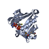
| Title | Crystal structure of Escherichia coli PliG in complex with Atlantic salmon g-type lysozyme | ||||||
 Components Components |
| ||||||
 Keywords Keywords | HYDROLASE/HYDROLASE INHIBITOR / hydrolase inhibitor /  lysozyme / HYDROLASE-HYDROLASE INHIBITOR complex lysozyme / HYDROLASE-HYDROLASE INHIBITOR complex | ||||||
| Function / homology |  Function and homology information Function and homology informationlysozyme inhibitor activity / peptidoglycan catabolic process / lysozyme / lysozyme activity / killing of cells of another organism / periplasmic space / defense response to bacterium / cytosolSimilarity search - Function | ||||||
| Biological species |  Salmo salar (Atlantic salmon) Salmo salar (Atlantic salmon) Escherichia coli (E. coli) Escherichia coli (E. coli) | ||||||
| Method | X-RAY DIFFRACTION / SYNCHROTRON / MOLECULAR REPLACEMENT / Resolution: 0.95 Å | ||||||
 Authors Authors | Leysen, S. / Vanderkelen, L. / Weeks, S.D. / Michiels, C.W. / Strelkov, S.V. | ||||||
 Citation Citation | Journal: Cell.Mol.Life Sci. / Year: 2013 Title: Structural basis of bacterial defense against g-type lysozyme-based innate immunity. Authors: Leysen, S. / Vanderkelen, L. / Weeks, S.D. / Michiels, C.W. / Strelkov, S.V. | ||||||
| History |
|
- Structure visualization
| Structure viewer | Molecule: MolmilJmol/JSmol |
|---|
- Downloads & links
-Download
| PDBx/mmCIF format | 4g9s.cif.gz | 215.5 KB | Display | PDBx/mmCIF format |
|---|---|---|---|---|
| PDB format | pdb4g9s.ent.gz | 177.3 KB | Display | PDB format |
| PDBx/mmJSON format | 4g9s.json.gz | Tree view | PDBx/mmJSON format | |
| Others |  Other downloads Other downloads |
-Validation report
| Arichive directory | https://data.pdbj.org/pub/pdb/validation_reports/g9/4g9sftp://data.pdbj.org/pub/pdb/validation_reports/g9/4g9s | HTTPS FTP |
|---|
-Related structure data


-Links
 PDBj
PDBj


- Assembly
| Deposited unit | 
| ||||||||
|---|---|---|---|---|---|---|---|---|---|
| 1 | 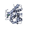
| ||||||||
| 2 | 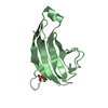
| ||||||||
| Unit cell |
|
-Components
| #1: Protein | Mass: 21094.729 Da / Num. of mol.: 1 / Fragment: UNP residues 22-200 / Mutation: A133V Source method: isolated from a genetically manipulated source Source: (gene. exp.) Salmo salar (Atlantic salmon) / Gene: lysG, LYG / Plasmid: pQE2 / Production host: Escherichia coli (E. coli) / Strain (production host): BL21(DE3) / References: UniProt: A6PZ97 |
|---|---|
| #2: Protein | Mass: 12538.815 Da / Num. of mol.: 1 / Fragment: UNP residues 23-133 Source method: isolated from a genetically manipulated source Source: (gene. exp.) Escherichia coli (E. coli) / Gene: pliG, ycgK / Plasmid: pETHSUL / Production host: Escherichia coli (E. coli) / Strain (production host): BL21(DE3) / References: UniProt: P76002 |
| #3: Chemical | ChemComp-CL / Chloride  Mass: 35.453 Da / Num. of mol.: 1 / Source method: obtained synthetically / Formula: Cl Mass: 35.453 Da / Num. of mol.: 1 / Source method: obtained synthetically / Formula: Cl |
| #4: Chemical | ChemComp-FLC / Citric acid  Mass: 189.100 Da / Num. of mol.: 1 / Source method: obtained synthetically / Formula: C6H5O7 Mass: 189.100 Da / Num. of mol.: 1 / Source method: obtained synthetically / Formula: C6H5O7 |
| #5: Water | ChemComp-HOH / Water Mass: 18.015 Da / Num. of mol.: 541 / Source method: isolated from a natural source / Formula: H2O Mass: 18.015 Da / Num. of mol.: 541 / Source method: isolated from a natural source / Formula: H2O |
-Experimental details
-Experiment
| Experiment | Method: X-RAY DIFFRACTION / Number of used crystals: 1 |
|---|
- Sample preparation
| Crystal | Density Matthews: 3.07 Å3/Da / Density % sol: 60 % |
|---|---|
| Crystal grow | Temperature: 293 K / Method: vapor diffusion, hanging drop / pH: 7 Details: 1.8M tri-ammonium citrate, pH 7, VAPOR DIFFUSION, HANGING DROP, temperature 293K |
-Data collection
| Diffraction |
| ||||||||||||||||||
|---|---|---|---|---|---|---|---|---|---|---|---|---|---|---|---|---|---|---|---|
| Diffraction source |
| ||||||||||||||||||
| Detector | Type: DECTRIS PILATUS 6M / Detector: PIXEL / Date: Feb 24, 2012 | ||||||||||||||||||
| Radiation |
| ||||||||||||||||||
| Radiation wavelength |
| ||||||||||||||||||
| Reflection | Resolution: 0.95→43.43 Å / Num. obs: 265542 / % possible obs: 98.5 % / Observed criterion σ(F): -3 / Observed criterion σ(I): 2 | ||||||||||||||||||
| Reflection shell | Resolution: 0.95→1 Å / Redundancy: 3 % / Mean I/σ(I) obs: 0.7 / Num. unique all: 36278 / % possible all: 92.4 |

- Processing
| Software |
| ||||||||||||||||||||||||||||||||||||||||||||||||||||||||||||||||||||||||||||||||||||||||||||||||||||||||||||||||||||||||||||||||||||||||||||
|---|---|---|---|---|---|---|---|---|---|---|---|---|---|---|---|---|---|---|---|---|---|---|---|---|---|---|---|---|---|---|---|---|---|---|---|---|---|---|---|---|---|---|---|---|---|---|---|---|---|---|---|---|---|---|---|---|---|---|---|---|---|---|---|---|---|---|---|---|---|---|---|---|---|---|---|---|---|---|---|---|---|---|---|---|---|---|---|---|---|---|---|---|---|---|---|---|---|---|---|---|---|---|---|---|---|---|---|---|---|---|---|---|---|---|---|---|---|---|---|---|---|---|---|---|---|---|---|---|---|---|---|---|---|---|---|---|---|---|---|---|---|
| Refinement | Method to determine structure: MOLECULAR REPLACEMENT Starting model: 3MGW AND 4DY3 Resolution: 0.95→43.267 Å / SU ML: 0.12 / σ(F): 1.33 / Phase error: 15.62 / Stereochemistry target values: ML
| ||||||||||||||||||||||||||||||||||||||||||||||||||||||||||||||||||||||||||||||||||||||||||||||||||||||||||||||||||||||||||||||||||||||||||||
| Solvent computation | Shrinkage radii: 0.9 Å / VDW probe radii: 1.11 Å / Solvent model: FLAT BULK SOLVENT MODEL | ||||||||||||||||||||||||||||||||||||||||||||||||||||||||||||||||||||||||||||||||||||||||||||||||||||||||||||||||||||||||||||||||||||||||||||
| Refinement step | Cycle: LAST / Resolution: 0.95→43.267 Å
| ||||||||||||||||||||||||||||||||||||||||||||||||||||||||||||||||||||||||||||||||||||||||||||||||||||||||||||||||||||||||||||||||||||||||||||
| Refine LS restraints |
| ||||||||||||||||||||||||||||||||||||||||||||||||||||||||||||||||||||||||||||||||||||||||||||||||||||||||||||||||||||||||||||||||||||||||||||
| LS refinement shell |
|
