 Movie
Movie Controller

[English] 日本語
 Yorodumi
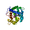
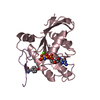
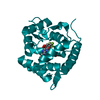
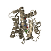
- PDB-4mrx: Crystal Structure of Y138F obelin mutant from Obelia longissima a... -
+ Open data

- Basic information
| Entry | Database: PDB / ID: 4mrx | ||||||
|---|---|---|---|---|---|---|---|
| Title | Crystal Structure of Y138F obelin mutant from Obelia longissima at 1.72 Angstrom resolution | ||||||
 Components Components | Obelin | ||||||
 Keywords Keywords |  LUMINESCENT PROTEIN / calcium binding / EF-hand LUMINESCENT PROTEIN / calcium binding / EF-hand | ||||||
| Function / homology |  Function and homology information Function and homology information | ||||||
| Biological species |  Obelia longissima (invertebrata) Obelia longissima (invertebrata) | ||||||
| Method | X-RAY DIFFRACTION / SYNCHROTRON / MOLECULAR REPLACEMENT / Resolution: 1.718 Å | ||||||
 Authors Authors | Natashin, P.V. / Ding, W. / Eremeeva, E.V. / Markova, S.V. / Lee, J. / Vysotski, E.S. / Liu, Z.J. | ||||||
 Citation Citation | Journal: Acta Crystallogr.,Sect.D / Year: 2014 Title: Structures of the Ca2+-regulated photoprotein obelin Y138F mutant before and after bioluminescence support the catalytic function of a water molecule in the reaction. Authors: Natashin, P.V. / Ding, W. / Eremeeva, E.V. / Markova, S.V. / Lee, J. / Vysotski, E.S. / Liu, Z.J. | ||||||
| History |
|
- Structure visualization
| Structure viewer | Molecule: MolmilJmol/JSmol |
|---|
- Downloads & links
-Download
| PDBx/mmCIF format | 4mrx.cif.gz | 107.8 KB | Display | PDBx/mmCIF format |
|---|---|---|---|---|
| PDB format | pdb4mrx.ent.gz | 82.8 KB | Display | PDB format |
| PDBx/mmJSON format | 4mrx.json.gz | Tree view | PDBx/mmJSON format | |
| Others |  Other downloads Other downloads |
-Validation report
| Arichive directory | https://data.pdbj.org/pub/pdb/validation_reports/mr/4mrxftp://data.pdbj.org/pub/pdb/validation_reports/mr/4mrx | HTTPS FTP |
|---|
-Related structure data
| Related structure data | 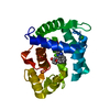 4mryC  1qv0S S: Starting model for refinement C: citing same article ( |
|---|---|
| Similar structure data |


-Links
 PDBj
PDBj- Assembly
| Deposited unit | 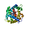
| ||||||||
|---|---|---|---|---|---|---|---|---|---|
| 1 |
| ||||||||
| Unit cell |
|
-Components
| #1: Protein | Mass: 22222.904 Da / Num. of mol.: 1 / Mutation: S2A, Y138F Source method: isolated from a genetically manipulated source Source: (gene. exp.) Obelia longissima (invertebrata) / Plasmid: pOL-138F / Production host:  Escherichia coli (E. coli) / Strain (production host): Bl21 Gold / References: UniProt: Q27709 Escherichia coli (E. coli) / Strain (production host): Bl21 Gold / References: UniProt: Q27709 |
|---|---|
| #2: Chemical | ChemComp-CZH / 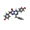  Mass: 455.462 Da / Num. of mol.: 1 / Source method: obtained synthetically / Formula: C26H21N3O5 Mass: 455.462 Da / Num. of mol.: 1 / Source method: obtained synthetically / Formula: C26H21N3O5 |
| #3: Water | ChemComp-HOH / Water Mass: 18.015 Da / Num. of mol.: 298 / Source method: isolated from a natural source / Formula: H2O Mass: 18.015 Da / Num. of mol.: 298 / Source method: isolated from a natural source / Formula: H2O |
-Experimental details
-Experiment
| Experiment | Method: X-RAY DIFFRACTION / Number of used crystals: 1 |
|---|
- Sample preparation
| Crystal | Density Matthews: 3.27 Å3/Da / Density % sol: 62.33 % |
|---|---|
| Crystal grow | Temperature: 277 K / Method: vapor diffusion, hanging drop / pH: 7 Details: 2.1M DL-Malic acid, pH 7.0, VAPOR DIFFUSION, HANGING DROP, temperature 277K |
-Data collection
| Diffraction | Mean temperature: 100 K |
|---|---|
| Diffraction source | Source: SYNCHROTRON / Site: SSRF  / Beamline: BL17U / Wavelength: 0.9789 Å / Beamline: BL17U / Wavelength: 0.9789 Å |
| Detector | Type: ADSC QUANTUM 315r / Detector: CCD / Date: Dec 25, 2012 |
| Radiation | Monochromator: Si 111 CHANNEL / Protocol: SINGLE WAVELENGTH / Monochromatic (M) / Laue (L): M / Scattering type: x-ray |
| Radiation wavelength | Wavelength: 0.9789 Å / Relative weight: 1 |
| Reflection | Resolution: 1.718→50 Å / Num. all: 30654 / Num. obs: 30654 / % possible obs: 100 % / Observed criterion σ(F): 2 / Observed criterion σ(I): 2 / Biso Wilson estimate: 21.28 Å2 |
| Reflection shell | Resolution: 1.72→1.78 Å / % possible all: 100 |
- Processing
| Software |
| |||||||||||||||||||||||||||||||||||||||||||||||||||||||||||||||||||||||||||||||||||||||||||||||||||||||||
|---|---|---|---|---|---|---|---|---|---|---|---|---|---|---|---|---|---|---|---|---|---|---|---|---|---|---|---|---|---|---|---|---|---|---|---|---|---|---|---|---|---|---|---|---|---|---|---|---|---|---|---|---|---|---|---|---|---|---|---|---|---|---|---|---|---|---|---|---|---|---|---|---|---|---|---|---|---|---|---|---|---|---|---|---|---|---|---|---|---|---|---|---|---|---|---|---|---|---|---|---|---|---|---|---|---|---|
| Refinement | Method to determine structure: MOLECULAR REPLACEMENT Starting model: 1QV0 Resolution: 1.718→37.357 Å / Occupancy max: 1 / Occupancy min: 0.22 / SU ML: 0.12 / σ(F): 1.35 / Phase error: 15.69 / Stereochemistry target values: ML
| |||||||||||||||||||||||||||||||||||||||||||||||||||||||||||||||||||||||||||||||||||||||||||||||||||||||||
| Solvent computation | Shrinkage radii: 0.9 Å / VDW probe radii: 1.11 Å / Solvent model: FLAT BULK SOLVENT MODEL | |||||||||||||||||||||||||||||||||||||||||||||||||||||||||||||||||||||||||||||||||||||||||||||||||||||||||
| Displacement parameters | Biso max: 101.72 Å2 / Biso mean: 26.3736 Å2 / Biso min: 11.49 Å2 | |||||||||||||||||||||||||||||||||||||||||||||||||||||||||||||||||||||||||||||||||||||||||||||||||||||||||
| Refinement step | Cycle: LAST / Resolution: 1.718→37.357 Å
| |||||||||||||||||||||||||||||||||||||||||||||||||||||||||||||||||||||||||||||||||||||||||||||||||||||||||
| Refine LS restraints |
| |||||||||||||||||||||||||||||||||||||||||||||||||||||||||||||||||||||||||||||||||||||||||||||||||||||||||
| LS refinement shell | Refine-ID: X-RAY DIFFRACTION / Total num. of bins used: 14
| |||||||||||||||||||||||||||||||||||||||||||||||||||||||||||||||||||||||||||||||||||||||||||||||||||||||||
| Refinement TLS params. | Method: refined / Origin x: 69.6173 Å / Origin y: 24.76 Å / Origin z: 22.9557 Å
| |||||||||||||||||||||||||||||||||||||||||||||||||||||||||||||||||||||||||||||||||||||||||||||||||||||||||
| Refinement TLS group | Selection details: ALL |
