 Movie
Movie Controller


+ Open data

- Basic information
| Entry | Database: PDB / ID: 4fzf | ||||||
|---|---|---|---|---|---|---|---|
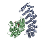
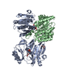
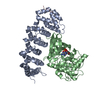
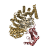
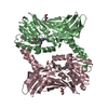
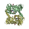
| Title | Crystal structure of MST4-MO25 complex with DKI | ||||||
 Components Components |
| ||||||
 Keywords Keywords |  SIGNALING PROTEIN/TRANSFERASE/INHIBITOR / Scaffold protein / Protein Ser/Thr kinase / ATP binding / SIGNALING PROTEIN-TRANSFERASE-INHIBITOR complex SIGNALING PROTEIN/TRANSFERASE/INHIBITOR / Scaffold protein / Protein Ser/Thr kinase / ATP binding / SIGNALING PROTEIN-TRANSFERASE-INHIBITOR complex | ||||||
| Function / homology |  Function and homology information Function and homology informationnegative regulation of potassium ion transmembrane transporter activity / microvillus assembly / negative regulation of potassium ion transmembrane transport / serine/threonine protein kinase complex / cellular hypotonic response / response to thyroid hormone / Energy dependent regulation of mTOR by LKB1-AMPK / vesicle membrane / Golgi-associated vesicle / protein kinase activator activity ...negative regulation of potassium ion transmembrane transporter activity / microvillus assembly / negative regulation of potassium ion transmembrane transport / serine/threonine protein kinase complex / cellular hypotonic response / response to thyroid hormone / Energy dependent regulation of mTOR by LKB1-AMPK / vesicle membrane / Golgi-associated vesicle / protein kinase activator activity / Apoptotic cleavage of cellular proteins / cellular response to starvation / protein serine/threonine kinase activator activity / negative regulation of cell migration / positive regulation of peptidyl-threonine phosphorylation / response to activity / cell periphery / positive regulation of protein serine/threonine kinase activity / kinase binding / Z disc / cellular response to oxidative stress / peptidyl-serine phosphorylation / regulation of apoptotic process / secretory granule lumen / ficolin-1-rich granule lumen / protein autophosphorylation / non-specific serine/threonine protein kinase / protein kinase activity / intracellular signal transduction / apical plasma membrane / protein phosphorylation / protein serine kinase activity / protein serine/threonine kinase activity / apoptotic process / Neutrophil degranulation / perinuclear region of cytoplasm / Golgi apparatus / magnesium ion binding / signal transduction / protein homodimerization activity / extracellular exosome / extracellular region / ATP binding / membrane / identical protein binding / cytosol / cytoplasmSimilarity search - Function | ||||||
| Biological species |  Homo sapiens (human) Homo sapiens (human) | ||||||
| Method | X-RAY DIFFRACTION / SYNCHROTRON / MOLECULAR REPLACEMENT / Resolution: 3.64 Å | ||||||
 Authors Authors | Shi, Z.B. / Zhou, Z.C. | ||||||
 Citation Citation | Journal: Structure / Year: 2013 Title: Structure of the MST4 in Complex with MO25 Provides Insights into Its Activation Mechanism Authors: Shi, Z. / Jiao, S. / Zhang, Z. / Ma, M. / Zhang, Z. / Chen, C. / Wang, K. / Wang, H. / Wang, W. / Zhang, L. / Zhao, Y. / Zhou, Z. | ||||||
| History |
|
- Structure visualization
| Structure viewer | Molecule: MolmilJmol/JSmol |
|---|
- Downloads & links
-Download
| PDBx/mmCIF format | 4fzf.cif.gz | 263.3 KB | Display | PDBx/mmCIF format |
|---|---|---|---|---|
| PDB format | pdb4fzf.ent.gz | 217 KB | Display | PDB format |
| PDBx/mmJSON format | 4fzf.json.gz | Tree view | PDBx/mmJSON format | |
| Others |  Other downloads Other downloads |
-Validation report
| Arichive directory | https://data.pdbj.org/pub/pdb/validation_reports/fz/4fzfftp://data.pdbj.org/pub/pdb/validation_reports/fz/4fzf | HTTPS FTP |
|---|
-Related structure data
| Related structure data |  4fzaC 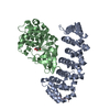 4fzdC  1uplS 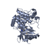 3ggfS C: citing same article ( S: Starting model for refinement |
|---|---|
| Similar structure data |


-Links
 PDBj
PDBj
- Assembly
| Deposited unit | 
| ||||||||
|---|---|---|---|---|---|---|---|---|---|
| 1 |
| ||||||||
| Unit cell |
|
-Components
| #1: Protein | Mass: 38311.098 Da / Num. of mol.: 1 / Fragment: Mo25-like, UNP residues 11-334 Source method: isolated from a genetically manipulated source Source: (gene. exp.) Homo sapiens (human) / Gene: CAB39, MO25, CGI-66 / Plasmid: HT-pET28a / Production host:  Escherichia coli (E. coli) / Strain (production host): BL21(DE3) / References: UniProt: Q9Y376 Escherichia coli (E. coli) / Strain (production host): BL21(DE3) / References: UniProt: Q9Y376 |
|---|---|
| #2: Protein | Mass: 31930.670 Da / Num. of mol.: 1 / Fragment: Kinase domain, UNP residues 18-297 / Mutation: D162A Source method: isolated from a genetically manipulated source Source: (gene. exp.) Homo sapiens (human) / Gene: MST4, MASK / Plasmid: HT-pET28a / Production host: Escherichia coli (E. coli) / Strain (production host): BL21(DE3)References: UniProt: Q9P289, non-specific serine/threonine protein kinase |
| #3: Chemical | ChemComp-DKI /   Mass: 425.436 Da / Num. of mol.: 1 / Source method: obtained synthetically / Formula: C15H13F2N7O2S2 Mass: 425.436 Da / Num. of mol.: 1 / Source method: obtained synthetically / Formula: C15H13F2N7O2S2 |
-Experimental details
-Experiment
| Experiment | Method: X-RAY DIFFRACTION / Number of used crystals: 1 |
|---|
- Sample preparation
| Crystal | Density Matthews: 4.07 Å3/Da / Density % sol: 69.8 % |
|---|---|
| Crystal grow | Temperature: 289 K / Method: vapor diffusion, hanging drop / pH: 8 Details: 0.1M Tris pH 8.0, 20% PEG 350 mme, VAPOR DIFFUSION, HANGING DROP, temperature 289K |
-Data collection
| Diffraction | Mean temperature: 100 K |
|---|---|
| Diffraction source | Source: SYNCHROTRON / Site: SSRF  / Beamline: BL17U / Wavelength: 0.97915 Å / Beamline: BL17U / Wavelength: 0.97915 Å |
| Detector | Type: ADSC QUANTUM 315 / Detector: CCD / Date: Sep 10, 2011 |
| Radiation | Monochromator: Si 111 CHANNEL / Protocol: SINGLE WAVELENGTH / Monochromatic (M) / Laue (L): M / Scattering type: x-ray |
| Radiation wavelength | Wavelength: 0.97915 Å / Relative weight: 1 |
| Reflection | Resolution: 3.64→50 Å / Num. all: 13410 / Num. obs: 13356 / % possible obs: 99.6 % / Observed criterion σ(F): 2 / Observed criterion σ(I): 2 / Redundancy: 12.8 % |
| Reflection shell | Resolution: 3.64→3.71 Å / Redundancy: 13.6 % / Mean I/σ(I) obs: 4.1 / Num. unique all: 651 / % possible all: 100 |
- Processing
| Software |
| |||||||||||||||||||||||||||||||||||||||||||||||||||||||||||||||||||||||||||
|---|---|---|---|---|---|---|---|---|---|---|---|---|---|---|---|---|---|---|---|---|---|---|---|---|---|---|---|---|---|---|---|---|---|---|---|---|---|---|---|---|---|---|---|---|---|---|---|---|---|---|---|---|---|---|---|---|---|---|---|---|---|---|---|---|---|---|---|---|---|---|---|---|---|---|---|---|
| Refinement | Method to determine structure: MOLECULAR REPLACEMENT Starting model: PDB ENTRY: 1UPL and 3GGF Resolution: 3.64→38.85 Å / Cor.coef. Fo:Fc: 0.911 / Cor.coef. Fo:Fc free: 0.831 / SU B: 73.766 / SU ML: 0.499 / Cross valid method: THROUGHOUT / σ(F): 2 / ESU R Free: 0.715 / Stereochemistry target values: MAXIMUM LIKELIHOOD / Details: HYDROGENS HAVE BEEN USED IF PRESENT IN THE INPUT
| |||||||||||||||||||||||||||||||||||||||||||||||||||||||||||||||||||||||||||
| Solvent computation | Ion probe radii: 0.8 Å / Shrinkage radii: 0.8 Å / VDW probe radii: 1.2 Å / Solvent model: MASK | |||||||||||||||||||||||||||||||||||||||||||||||||||||||||||||||||||||||||||
| Displacement parameters | Biso mean: 116.313 Å2 | |||||||||||||||||||||||||||||||||||||||||||||||||||||||||||||||||||||||||||
| Refinement step | Cycle: LAST / Resolution: 3.64→38.85 Å
| |||||||||||||||||||||||||||||||||||||||||||||||||||||||||||||||||||||||||||
| Refine LS restraints |
| |||||||||||||||||||||||||||||||||||||||||||||||||||||||||||||||||||||||||||
| LS refinement shell | Resolution: 3.644→3.739 Å / Total num. of bins used: 20
| |||||||||||||||||||||||||||||||||||||||||||||||||||||||||||||||||||||||||||
| Refinement TLS params. | Method: refined / Refine-ID: X-RAY DIFFRACTION
| |||||||||||||||||||||||||||||||||||||||||||||||||||||||||||||||||||||||||||
| Refinement TLS group |
|
