 Movie
Movie Controller

[English] 日本語
 Yorodumi
- PDB-4fsa: tRNA-Guanine Transglycosylase soaked with pyridyl-alkine-substitu... -
+ Open data

- Basic information
| Entry | Database: PDB / ID: 4fsa | ||||||
|---|---|---|---|---|---|---|---|
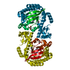
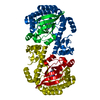
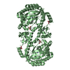
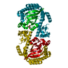
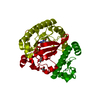
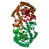
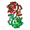
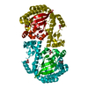
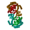
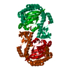
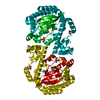
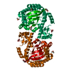
| Title | tRNA-Guanine Transglycosylase soaked with pyridyl-alkine-substituted lin-benzoguanine ligand | ||||||
 Components Components | Queuine tRNA-ribosyltransferase | ||||||
 Keywords Keywords | TRANSFERASE/TRANSFERASE inhibitor / TIM BARREL / GLYCOSYLTRANSFERASE / QUEUOSINE / BIOSYNTHESIS / TRANSFERASE / TRNA PROCESSING / TRNA / TRANSFERASE-TRANSFERASE inhibitor complex | ||||||
| Function / homology |  Function and homology information Function and homology informationtRNA-guanosine34 preQ1 transglycosylase / tRNA wobble guanine modification / tRNA-guanosine(34) queuine transglycosylase activity / tRNA-guanine transglycosylation / queuosine biosynthetic process / metal ion binding / cytosolSimilarity search - Function | ||||||
| Biological species |  Zymomonas mobilis subsp. mobilis (bacteria) Zymomonas mobilis subsp. mobilis (bacteria) | ||||||
| Method | X-RAY DIFFRACTION / SYNCHROTRON / MOLECULAR REPLACEMENT / Resolution: 1.62 Å | ||||||
 Authors Authors | Immekus, F. / Klebe, G. | ||||||
 Citation Citation | Journal: To be Published Title: Studies on TGT homodimer interface Authors: Immekus, F. / Barandun, L.J. / Betz, M. / Debaene, F. / Petiot, S. / Sanglier-Cianferani, S. / Diederich, F. / Klebe, G. | ||||||
| History |
|
- Structure visualization
| Structure viewer | Molecule: MolmilJmol/JSmol |
|---|
- Downloads & links
-Download
| PDBx/mmCIF format | 4fsa.cif.gz | 88.3 KB | Display | PDBx/mmCIF format |
|---|---|---|---|---|
| PDB format | pdb4fsa.ent.gz | 62.9 KB | Display | PDB format |
| PDBx/mmJSON format | 4fsa.json.gz | Tree view | PDBx/mmJSON format | |
| Others |  Other downloads Other downloads |
-Validation report
| Arichive directory | https://data.pdbj.org/pub/pdb/validation_reports/fs/4fsaftp://data.pdbj.org/pub/pdb/validation_reports/fs/4fsa | HTTPS FTP |
|---|
-Related structure data
| Related structure data | 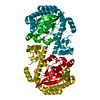 4fpsC 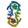 4fr1C  4fr6C 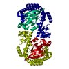 1p0dS C: citing same article ( S: Starting model for refinement |
|---|---|
| Similar structure data |


-Links
 PDBj
PDBj- Assembly
| Deposited unit | 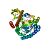
| ||||||||
|---|---|---|---|---|---|---|---|---|---|
| 1 | 
| ||||||||
| Unit cell |
| ||||||||
| Details | HOMODIMER |
-Components
| #1: Protein | / Guanine insertion enzyme / tRNA-guanine transglycosylase Mass: 42925.703 Da / Num. of mol.: 1 Source method: isolated from a genetically manipulated source Source: (gene. exp.) Zymomonas mobilis subsp. mobilis (bacteria)Strain: ATCC 31821 / ZM4 / CP4 / Gene: tgt, ZMO0363 / Plasmid: PET9D / Production host: Escherichia coli (E. coli) / Strain (production host): BL21(DE3)References: UniProt: P28720, tRNA-guanosine34 preQ1 transglycosylase |
|---|---|
| #2: Chemical | ChemComp-ZN /   Mass: 65.409 Da / Num. of mol.: 1 / Source method: obtained synthetically / Formula: Zn Mass: 65.409 Da / Num. of mol.: 1 / Source method: obtained synthetically / Formula: Zn |
| #3: Chemical | ChemComp-0V3 /   Mass: 470.569 Da / Num. of mol.: 1 / Source method: obtained synthetically / Formula: C26H30N8O Mass: 470.569 Da / Num. of mol.: 1 / Source method: obtained synthetically / Formula: C26H30N8O |
| #4: Chemical | ChemComp-GOL / Glycerol  Mass: 92.094 Da / Num. of mol.: 1 / Source method: obtained synthetically / Formula: C3H8O3 Mass: 92.094 Da / Num. of mol.: 1 / Source method: obtained synthetically / Formula: C3H8O3 |
| #5: Water | ChemComp-HOH / Water Mass: 18.015 Da / Num. of mol.: 242 / Source method: isolated from a natural source / Formula: H2O Mass: 18.015 Da / Num. of mol.: 242 / Source method: isolated from a natural source / Formula: H2O |
-Experimental details
-Experiment
| Experiment | Method: X-RAY DIFFRACTION / Number of used crystals: 1 |
|---|
- Sample preparation
| Crystal | Density Matthews: 2.38 Å3/Da / Density % sol: 49.21 % |
|---|---|
| Crystal grow | Temperature: 288 K / Method: vapor diffusion, hanging drop / pH: 5.5 Details: 100 MM MES, 1MM DTT, 10% DMSO, 13% PEG8000, pH 5.5, VAPOR DIFFUSION, HANGING DROP, temperature 288K |
-Data collection
| Diffraction | Mean temperature: 100 K |
|---|---|
| Diffraction source | Source: SYNCHROTRON / Site: BESSY  / Beamline: 14.2 / Wavelength: 0.91841 Å / Beamline: 14.2 / Wavelength: 0.91841 Å |
| Detector | Type: RAYONIX MX-225 / Detector: CCD / Date: Apr 24, 2012 / Details: Rh-coated silicon with indirect water cooling |
| Radiation | Monochromator: Double crystal / Protocol: SINGLE WAVELENGTH / Monochromatic (M) / Laue (L): M / Scattering type: x-ray |
| Radiation wavelength | Wavelength: 0.91841 Å / Relative weight: 1 |
| Reflection | Resolution: 1.62→50 Å / Num. all: 51849 / Num. obs: 51849 / % possible obs: 99.5 % / Redundancy: 2.9 % / Biso Wilson estimate: 18.34 Å2 / Rsym value: 0.05 / Net I/σ(I): 19.16 |
| Reflection shell | Resolution: 1.62→1.65 Å / Redundancy: 2.9 % / Mean I/σ(I) obs: 3.35 / Num. unique all: 2587 / Rsym value: 0.346 / % possible all: 99.6 |
- Processing
| Software |
| |||||||||||||||||||||||||||||||||
|---|---|---|---|---|---|---|---|---|---|---|---|---|---|---|---|---|---|---|---|---|---|---|---|---|---|---|---|---|---|---|---|---|---|---|
| Refinement | Method to determine structure: MOLECULAR REPLACEMENT Starting model: pdb entry 1P0D Resolution: 1.62→50 Å / Num. parameters: 11603 / Num. restraintsaints: 10970 / Cross valid method: FREE R / σ(F): 0 / σ(I): 0 / Stereochemistry target values: ENGH AND HUBER Details: ANISOTROPIC SCALING APPLIED BY THE METHOD OF PARKIN, MOEZZI & HOPE, J.APPL.CRYST.28(1995)53-56
| |||||||||||||||||||||||||||||||||
| Refine analyze | Num. disordered residues: 5 / Occupancy sum hydrogen: 2540 / Occupancy sum non hydrogen: 2877 | |||||||||||||||||||||||||||||||||
| Refinement step | Cycle: LAST / Resolution: 1.62→50 Å
| |||||||||||||||||||||||||||||||||
| Refine LS restraints |
|
