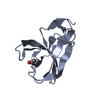
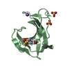
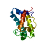
- PDB-4fjo: Structure of the Rev1 CTD-Rev3/7-Pol kappa RIR complex -
+
Open data
ID or keywords:
Loading...
-
Basic information
Entry
Database: PDB / ID: 4fjo
Title
Structure of the Rev1 CTD-Rev3/7-Pol kappa RIR complex
Components
(DNA polymerase ...) x 2
DNA repair protein REV1
Mitotic spindle assembly checkpoint protein MAD2B
Keywords
TRANSFERASE/DNA BINDING PROTEIN / Translesion Synthesis / TRANSFERASE -DNA BINDING PROTEIN complex / TRANSFERASE-DNA BINDING PROTEIN complex
Function / homology
Function and homology information
: / Gap-filling DNA repair synthesis and ligation in GG-NER / Translesion synthesis by REV1 / Translesion synthesis by POLK / Translesion synthesis by POLI / HDR through Homologous Recombination (HRR) / somatic diversification of immunoglobulins involved in immune response / DNA damage response, signal transduction resulting in transcription / Termination of translesion DNA synthesis / Dual Incision in GG-NER ...: / Gap-filling DNA repair synthesis and ligation in GG-NER / Translesion synthesis by REV1 / Translesion synthesis by POLK / Translesion synthesis by POLI / HDR through Homologous Recombination (HRR) / somatic diversification of immunoglobulins involved in immune response / DNA damage response, signal transduction resulting in transcription / Termination of translesion DNA synthesis / Dual Incision in GG-NER / Dual incision in TC-NER / Gap-filling DNA repair synthesis and ligation in TC-NER / deoxycytidyl transferase activity / telomere maintenance in response to DNA damage / zeta DNA polymerase complex / anaphase-promoting complex / positive regulation of isotype switching / positive regulation of extracellular matrix assembly / negative regulation of transcription by competitive promoter binding / negative regulation of cell-cell adhesion mediated by cadherin / negative regulation of epithelial to mesenchymal transition / JUN kinase binding / nucleotide-excision repair, DNA gap filling / negative regulation of ubiquitin protein ligase activity / error-free translesion synthesis / positive regulation of double-strand break repair via nonhomologous end joining / site of DNA damage / negative regulation of double-strand break repair via homologous recombination / error-prone translesion synthesis / positive regulation of epithelial to mesenchymal transition / response to UV / actin filament organization / regulation of cell growth / double-strand break repair via homologous recombination / negative regulation of canonical Wnt signaling pathway / negative regulation of protein catabolic process / spindle / cellular response to UV / double-strand break repair / Transferases; Transferring phosphorus-containing groups; Nucleotidyltransferases / positive regulation of peptidyl-serine phosphorylation / site of double-strand break / 4 iron, 4 sulfur cluster binding / RNA polymerase II-specific DNA-binding transcription factor binding / DNA replication / damaged DNA binding / DNA-directed DNA polymerase / molecular adaptor activity / DNA-directed DNA polymerase activity / nuclear body / cell cycle / cell division / nucleotide binding / DNA repair / DNA damage response / chromatin / positive regulation of gene expression / nucleolus / positive regulation of DNA-templated transcription / negative regulation of transcription by RNA polymerase II / DNA binding / nucleoplasm / metal ion binding / nucleus / cytoplasm Similarity search - Function
DNA repair protein Rev1, C-terminal domain / DNA repair protein Rev1, C-terminal / Rev1, C-terminal domain superfamily / : / DNA repair protein REV1 C-terminal domain / Domain of unknown function DUF4683 / Domain of unknown function (DUF4683) / Cell Cycle, Spindle Assembly Checkpoint Protein; Chain A / HORMA domain / DNA polymerase zeta catalytic subunit ...DNA repair protein Rev1, C-terminal domain / DNA repair protein Rev1, C-terminal / Rev1, C-terminal domain superfamily / : / DNA repair protein REV1 C-terminal domain / Domain of unknown function DUF4683 / Domain of unknown function (DUF4683) / Cell Cycle, Spindle Assembly Checkpoint Protein; Chain A / HORMA domain / DNA polymerase zeta catalytic subunit / HUWE1/Rev1, ubiquitin binding region / Ubiquitin binding region / Rad18-like CCHC zinc finger / DNA repair protein Rev1 / C4-type zinc-finger of DNA polymerase delta / C4-type zinc-finger of DNA polymerase delta / Mad2-like / HORMA domain / HORMA domain / HORMA domain profile. / HORMA domain superfamily / DNA polymerase IV / DNApol eta/Rev1, HhH motif / DNA polymerase family B, thumb domain / Rad18, zinc finger UBZ4-type / Zinc finger UBZ4-type profile. / DNA polymerase family B signature. / DNA-directed DNA polymerase, family B, conserved site / DNA polymerase family B / DNA polymerase family B, exonuclease domain / DNA-directed DNA polymerase, family B, exonuclease domain / DNA-directed DNA polymerase, family B, multifunctional domain / DNA polymerase, palm domain superfamily / DNA polymerase type-B family / DNA-directed DNA polymerase, family B / BRCA1 C Terminus (BRCT) domain / DNA polymerase, Y-family, little finger domain / impB/mucB/samB family C-terminal domain / UmuC domain / DNA polymerase, Y-family, little finger domain superfamily / impB/mucB/samB family / UmuC domain profile. / breast cancer carboxy-terminal domain / BRCT domain profile. / BRCT domain / BRCT domain superfamily / Methane Monooxygenase Hydroxylase; Chain G, domain 1 / Ribonuclease H superfamily / Ribonuclease H-like superfamily / Reverse transcriptase/Diguanylate cyclase domain / DNA/RNA polymerase superfamily / Up-down Bundle / 2-Layer Sandwich / Mainly Alpha / Alpha Beta Similarity search - Domain/homology
PHOSPHATE ION / DNA polymerase zeta catalytic subunit / DNA repair protein REV1 / Mitotic spindle assembly checkpoint protein MAD2B / DNA polymerase kappa Similarity search - Component
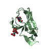
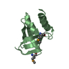
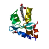
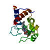
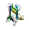
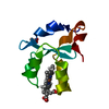
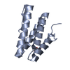
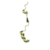
A: DNA repair protein REV1 B: DNA polymerase kappa C: Mitotic spindle assembly checkpoint protein MAD2B D: DNA polymerase zeta catalytic subunit hetero molecules
Mass: 18.015 Da / Num. of mol.: 131 / Source method: isolated from a natural source / Formula: H2O
-
Experimental details
-
Experiment
Experiment
Method: X-RAY DIFFRACTION / Number of used crystals: 1
-
Sample preparation
Crystal
Density Matthews: 4.69 Å3/Da / Density % sol: 73.79 %
Crystal grow
Temperature: 277 K / Method: vapor diffusion, sitting drop / pH: 8.5 Details: 0.1 M Tris pH8.5, 1.5 M Ammonium dihydrogen phosphate., VAPOR DIFFUSION, SITTING DROP, temperature 277K
In the structure databanks used in Yorodumi, some data are registered as the other names, "COVID-19 virus" and "2019-nCoV". Here are the details of the virus and the list of structure data.
Jan 31, 2019. EMDB accession codes are about to change! (news from PDBe EMDB page)
EMDB accession codes are about to change! (news from PDBe EMDB page)
The allocation of 4 digits for EMDB accession codes will soon come to an end. Whilst these codes will remain in use, new EMDB accession codes will include an additional digit and will expand incrementally as the available range of codes is exhausted. The current 4-digit format prefixed with “EMD-” (i.e. EMD-XXXX) will advance to a 5-digit format (i.e. EMD-XXXXX), and so on. It is currently estimated that the 4-digit codes will be depleted around Spring 2019, at which point the 5-digit format will come into force.
The EM Navigator/Yorodumi systems omit the EMD- prefix.
Related info.:Q: What is EMD? / ID/Accession-code notation in Yorodumi/EM Navigator
Yorodumi is a browser for structure data from EMDB, PDB, SASBDB, etc.
This page is also the successor to EM Navigator detail page, and also detail information page/front-end page for Omokage search.
The word "yorodu" (or yorozu) is an old Japanese word meaning "ten thousand". "mi" (miru) is to see.
Related info.:EMDB / PDB / SASBDB / Comparison of 3 databanks / Yorodumi Search / Aug 31, 2016. New EM Navigator & Yorodumi / Yorodumi Papers / Jmol/JSmol / Function and homology information / Changes in new EM Navigator and Yorodumi
 Movie
Movie Controller


 Open data
 Basic information
 Components
 Keywords
 Translesion Synthesis / TRANSFERASE -DNA BINDING PROTEIN complex / TRANSFERASE-DNA BINDING PROTEIN complex
 Function and homology information
 Authors
 Citation
 Structure visualization
 Downloads & links
 Other downloads
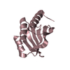
 PDBj
PDBj


 Assembly


 Mass: 92.094 Da / Num. of mol.: 1 / Source method: obtained synthetically / Formula: C3H8O3
 Mass: 94.971 Da / Num. of mol.: 8 / Source method: obtained synthetically / Formula: PO4
 Sample preparation
 / Beamline: 22-BM / Wavelength: 1 Å
 Processing