 Movie
Movie Controller

[English] 日本語
 Yorodumi
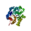
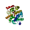
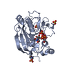
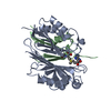
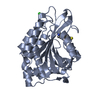
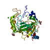
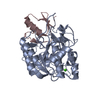
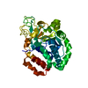
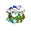
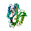
- PDB-4fai: Crystal structure of mitochondrial isoform of glutaminyl cyclase ... -
+ Open data

- Basic information
| Entry | Database: PDB / ID: 4fai | ||||||
|---|---|---|---|---|---|---|---|
| Title | Crystal structure of mitochondrial isoform of glutaminyl cyclase from Drosophila melanogaster | ||||||
 Components Components | CG5976, isoform B | ||||||
 Keywords Keywords |  TRANSFERASE / HYDROLASE / alpha/beta hydrolase / pGlu formation / PE / Alzheimer's Disease / pyroglutamate / pGlu-amyloid TRANSFERASE / HYDROLASE / alpha/beta hydrolase / pGlu formation / PE / Alzheimer's Disease / pyroglutamate / pGlu-amyloid | ||||||
| Function / homology |  Function and homology information Function and homology informationpeptidyl-pyroglutamic acid biosynthetic process, using glutaminyl-peptide cyclotransferase / glutaminyl-peptide cyclotransferase / glutaminyl-peptide cyclotransferase activity / Neutrophil degranulation / mitochondrion / zinc ion binding / extracellular regionSimilarity search - Function | ||||||
| Biological species |  Drosophila melanogaster (fruit fly) Drosophila melanogaster (fruit fly) | ||||||
| Method | X-RAY DIFFRACTION / SYNCHROTRON / MOLECULAR REPLACEMENT / Resolution: 1.65 Å | ||||||
 Authors Authors | Kolenko, P. / Koch, B. / Ruiz-Carilo, D. / Stubbs, M.T. | ||||||
 Citation Citation | Journal: Biochemistry / Year: 2012 Title: Crystal Structures of Glutaminyl Cyclases (QCs) from Drosophila melanogaster Reveal Active Site Conservation between Insect and Mammalian QCs. Authors: Koch, B. / Kolenko, P. / Buchholz, M. / Ruiz Carrillo, D. / Parthier, C. / Wermann, M. / Rahfeld, J.U. / Reuter, G. / Schilling, S. / Stubbs, M.T. / Demuth, H.U. | ||||||
| History |
|
- Structure visualization
| Structure viewer | Molecule: MolmilJmol/JSmol |
|---|
- Downloads & links
-Download
| PDBx/mmCIF format | 4fai.cif.gz | 147.9 KB | Display | PDBx/mmCIF format |
|---|---|---|---|---|
| PDB format | pdb4fai.ent.gz | 114.3 KB | Display | PDB format |
| PDBx/mmJSON format | 4fai.json.gz | Tree view | PDBx/mmJSON format | |
| Others |  Other downloads Other downloads |
-Validation report
| Arichive directory | https://data.pdbj.org/pub/pdb/validation_reports/fa/4faiftp://data.pdbj.org/pub/pdb/validation_reports/fa/4fai | HTTPS FTP |
|---|
-Related structure data
| Related structure data | 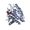 4f9uC 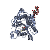 4f9vSC 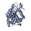 4fbeC C: citing same article ( S: Starting model for refinement |
|---|---|
| Similar structure data |
-Links
 PDBj
PDBj


- Assembly
| Deposited unit | 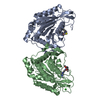
| ||||||||
|---|---|---|---|---|---|---|---|---|---|
| 1 | 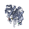
| ||||||||
| 2 | 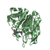
| ||||||||
| Unit cell |
|
-Components
| #1: Protein | Mass: 37930.301 Da / Num. of mol.: 2 / Fragment: UNP residues 32-354 Source method: isolated from a genetically manipulated source Source: (gene. exp.) Drosophila melanogaster (fruit fly) / Gene: isoQC, CG5976, Dmel_CG5976 / Plasmid: PQE31 / Production host:  Escherichia coli (E. coli) / Strain (production host): M15 Escherichia coli (E. coli) / Strain (production host): M15References: UniProt: Q86PD7, glutaminyl-peptide cyclotransferase, Hydrolases; Acting on peptide bonds (peptidases)#2: Chemical |   Mass: 65.409 Da / Num. of mol.: 2 / Source method: obtained synthetically / Formula: Zn Mass: 65.409 Da / Num. of mol.: 2 / Source method: obtained synthetically / Formula: Zn#3: Chemical | 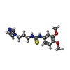  Mass: 320.410 Da / Num. of mol.: 2 / Source method: obtained synthetically / Formula: C15H20N4O2S Mass: 320.410 Da / Num. of mol.: 2 / Source method: obtained synthetically / Formula: C15H20N4O2S#4: Chemical | Chloride  Mass: 35.453 Da / Num. of mol.: 2 / Source method: obtained synthetically / Formula: Cl Mass: 35.453 Da / Num. of mol.: 2 / Source method: obtained synthetically / Formula: Cl#5: Water | ChemComp-HOH / | Water Mass: 18.015 Da / Num. of mol.: 446 / Source method: isolated from a natural source / Formula: H2O Mass: 18.015 Da / Num. of mol.: 446 / Source method: isolated from a natural source / Formula: H2O |
|---|
-Experimental details
-Experiment
| Experiment | Method: X-RAY DIFFRACTION / Number of used crystals: 1 |
|---|
- Sample preparation
| Crystal | Density Matthews: 2.02 Å3/Da / Density % sol: 39.13 % |
|---|---|
| Crystal grow | Temperature: 294 K / Method: vapor diffusion, hanging drop / pH: 8.5 Details: 30% PEG4000, 0.2 M magnesium chloride, 2 mM PQ50, 0.1 M Tris-HCl, pH 8.5, VAPOR DIFFUSION, HANGING DROP, temperature 294K |
-Data collection
| Diffraction | Mean temperature: 100 K |
|---|---|
| Diffraction source | Source: SYNCHROTRON / Site: BESSY  / Beamline: 14.1 / Wavelength: 0.91841 Å / Beamline: 14.1 / Wavelength: 0.91841 Å |
| Detector | Type: RAYONIX MX-225 / Detector: CCD / Date: Jul 10, 2010 / Details: mirrors |
| Radiation | Monochromator: double crystal Si(111) / Protocol: SINGLE WAVELENGTH / Monochromatic (M) / Laue (L): M / Scattering type: x-ray |
| Radiation wavelength | Wavelength: 0.91841 Å / Relative weight: 1 |
| Reflection | Resolution: 1.65→71.969 Å / Num. all: 69017 / Num. obs: 69017 / % possible obs: 97 % / Observed criterion σ(F): 0 / Observed criterion σ(I): -3.7 / Redundancy: 4 % / Biso Wilson estimate: 19 Å2 / Rmerge(I) obs: 0.05 / Net I/σ(I): 14.1 |
| Reflection shell | Resolution: 1.65→1.74 Å / Redundancy: 4 % / Rmerge(I) obs: 0.489 / Mean I/σ(I) obs: 2.3 / Num. unique all: 9906 / % possible all: 95 |
- Processing
| Software |
| ||||||||||||||||||||||||||||||||||||||||||||||||||||||||||||
|---|---|---|---|---|---|---|---|---|---|---|---|---|---|---|---|---|---|---|---|---|---|---|---|---|---|---|---|---|---|---|---|---|---|---|---|---|---|---|---|---|---|---|---|---|---|---|---|---|---|---|---|---|---|---|---|---|---|---|---|---|---|
| Refinement | Method to determine structure: MOLECULAR REPLACEMENT Starting model: PDB ENTRY 4F9V Resolution: 1.65→71.969 Å / Cor.coef. Fo:Fc: 0.965 / SU B: 1.603 / SU ML: 0.054 / Cross valid method: THROUGHOUT / ESU R: 0.098 / Stereochemistry target values: MAXIMUM LIKELIHOOD Details: HYDROGENS HAVE BEEN ADDED IN THE RIDING POSITIONS U VALUES : REFINED INDIVIDUALLY
| ||||||||||||||||||||||||||||||||||||||||||||||||||||||||||||
| Solvent computation | Ion probe radii: 0.8 Å / Shrinkage radii: 0.8 Å / VDW probe radii: 1.2 Å / Solvent model: MASK | ||||||||||||||||||||||||||||||||||||||||||||||||||||||||||||
| Displacement parameters | Biso mean: 17.439 Å2
| ||||||||||||||||||||||||||||||||||||||||||||||||||||||||||||
| Refinement step | Cycle: LAST / Resolution: 1.65→71.969 Å
| ||||||||||||||||||||||||||||||||||||||||||||||||||||||||||||
| Refine LS restraints |
| ||||||||||||||||||||||||||||||||||||||||||||||||||||||||||||
| LS refinement shell | Resolution: 1.65→1.693 Å / Total num. of bins used: 20
|
