 Movie
Movie Controller

[English] 日本語
 Yorodumi
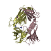
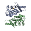
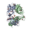
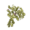
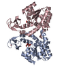
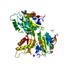
- PDB-4f4j: Conversion of the enzyme guanylate kinase into a mitotic spindle ... -
+ Open data

- Basic information
| Entry | Database: PDB / ID: 4f4j | |||||||||
|---|---|---|---|---|---|---|---|---|---|---|
| Title | Conversion of the enzyme guanylate kinase into a mitotic spindle orienting protein by a single mutation that inhibits gmp- induced closing | |||||||||
 Components Components | Guanylate kinase | |||||||||
 Keywords Keywords | TRANSFERASE / Phosphotransferase / GMP and ATP | |||||||||
| Function / homology |  Function and homology information Function and homology informationAzathioprine ADME / GDP biosynthetic process / Interconversion of nucleotide di- and triphosphates / guanylate kinase / purine nucleotide metabolic process / guanylate kinase activity / phosphorylation / ATP binding / nucleus / cytosol / cytoplasmSimilarity search - Function | |||||||||
| Biological species |  Saccharomyces cerevisiae (brewer's yeast) Saccharomyces cerevisiae (brewer's yeast) | |||||||||
| Method | X-RAY DIFFRACTION / SYNCHROTRON / MOLECULAR REPLACEMENT / Resolution: 2.45 Å | |||||||||
 Authors Authors | Johnston, C.A. / Whitney, D. / Volkman, B.F. / Prehoda, K.E. | |||||||||
 Citation Citation | Journal: Proc.Natl.Acad.Sci.USA / Year: 2011 Title: Conversion of the enzyme guanylate kinase into a mitotic-spindle orienting protein by a single mutation that inhibits GMP-induced closing. Authors: Johnston, C.A. / Whitney, D.S. / Volkman, B.F. / Doe, C.Q. / Prehoda, K.E. | |||||||||
| History |
|
- Structure visualization
| Structure viewer | Molecule: MolmilJmol/JSmol |
|---|
- Downloads & links
-Download
| PDBx/mmCIF format | 4f4j.cif.gz | 157.4 KB | Display | PDBx/mmCIF format |
|---|---|---|---|---|
| PDB format | pdb4f4j.ent.gz | 126.2 KB | Display | PDB format |
| PDBx/mmJSON format | 4f4j.json.gz | Tree view | PDBx/mmJSON format | |
| Others |  Other downloads Other downloads |
-Validation report
| Arichive directory | https://data.pdbj.org/pub/pdb/validation_reports/f4/4f4jftp://data.pdbj.org/pub/pdb/validation_reports/f4/4f4j | HTTPS FTP |
|---|
-Related structure data
| Similar structure data |
|---|


-Links
 PDBj
PDBj- Assembly
| Deposited unit | 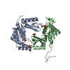
| ||||||||
|---|---|---|---|---|---|---|---|---|---|
| 1 |
| ||||||||
| 2 | 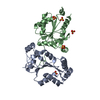
| ||||||||
| Unit cell |
|
-Components
| #1: Protein | / GMP kinase Mass: 22419.205 Da / Num. of mol.: 2 / Fragment: GMP kinase (unp residues 1-187) / Mutation: S35P Source method: isolated from a genetically manipulated source Source: (gene. exp.) Saccharomyces cerevisiae (brewer's yeast)Strain: ATCC 204508 / S288c / Gene: GUK1, YDR454C, D9461.39 / Production host:  Escherichia coli (E. coli) / References: UniProt: P15454, guanylate kinase Escherichia coli (E. coli) / References: UniProt: P15454, guanylate kinase#2: Chemical | ChemComp-SO4 / Sulfate  Mass: 96.063 Da / Num. of mol.: 5 / Source method: obtained synthetically / Formula: SO4 Mass: 96.063 Da / Num. of mol.: 5 / Source method: obtained synthetically / Formula: SO4#3: Water | ChemComp-HOH / | Water Mass: 18.015 Da / Num. of mol.: 91 / Source method: isolated from a natural source / Formula: H2O Mass: 18.015 Da / Num. of mol.: 91 / Source method: isolated from a natural source / Formula: H2O |
|---|
-Experimental details
-Experiment
| Experiment | Method: X-RAY DIFFRACTION / Number of used crystals: 1 |
|---|
- Sample preparation
| Crystal | Density Matthews: 3.92 Å3/Da / Density % sol: 68.64 % |
|---|---|
| Crystal grow | Temperature: 288 K / Method: vapor diffusion, sitting drop / pH: 7.5 Details: 1.6M LiSO4, 0.1M HEPES, pH 7.5, VAPOR DIFFUSION, SITTING DROP, temperature 288K |
-Data collection
| Diffraction | Mean temperature: 100 K |
|---|---|
| Diffraction source | Source: SYNCHROTRON / Site: ALS  / Beamline: 8.2.1 / Wavelength: 1 Å / Beamline: 8.2.1 / Wavelength: 1 Å |
| Detector | Type: ADSC QUANTUM 315r / Detector: CCD / Date: Jun 16, 2011 |
| Radiation | Monochromator: Double crystal, Si(111) / Protocol: SINGLE WAVELENGTH / Monochromatic (M) / Laue (L): M / Scattering type: x-ray |
| Radiation wavelength | Wavelength: 1 Å / Relative weight: 1 |
| Reflection | Resolution: 2.45→50 Å / Num. all: 25356 / Num. obs: 24012 / % possible obs: 98 % / Observed criterion σ(F): 2 / Observed criterion σ(I): 2 |
- Processing
| Software |
| |||||||||||||||||||||||||||||||||||||||||||||||||||||||||||||||||||||||||||
|---|---|---|---|---|---|---|---|---|---|---|---|---|---|---|---|---|---|---|---|---|---|---|---|---|---|---|---|---|---|---|---|---|---|---|---|---|---|---|---|---|---|---|---|---|---|---|---|---|---|---|---|---|---|---|---|---|---|---|---|---|---|---|---|---|---|---|---|---|---|---|---|---|---|---|---|---|
| Refinement | Method to determine structure: MOLECULAR REPLACEMENT / Resolution: 2.45→50 Å / Cor.coef. Fo:Fc: 0.937 / Cor.coef. Fo:Fc free: 0.914 / SU B: 14.769 / SU ML: 0.155 / Cross valid method: THROUGHOUT / σ(F): 3 / ESU R: 0.258 / ESU R Free: 0.221 / Stereochemistry target values: MAXIMUM LIKELIHOOD / Details: HYDROGENS HAVE BEEN ADDED IN THE RIDING POSITIONS
| |||||||||||||||||||||||||||||||||||||||||||||||||||||||||||||||||||||||||||
| Solvent computation | Ion probe radii: 0.8 Å / Shrinkage radii: 0.8 Å / VDW probe radii: 1.4 Å / Solvent model: MASK | |||||||||||||||||||||||||||||||||||||||||||||||||||||||||||||||||||||||||||
| Displacement parameters | Biso mean: 41.682 Å2
| |||||||||||||||||||||||||||||||||||||||||||||||||||||||||||||||||||||||||||
| Refinement step | Cycle: LAST / Resolution: 2.45→50 Å
| |||||||||||||||||||||||||||||||||||||||||||||||||||||||||||||||||||||||||||
| Refine LS restraints |
| |||||||||||||||||||||||||||||||||||||||||||||||||||||||||||||||||||||||||||
| LS refinement shell | Resolution: 2.45→2.514 Å / Total num. of bins used: 20
| |||||||||||||||||||||||||||||||||||||||||||||||||||||||||||||||||||||||||||
| Refinement TLS params. | Method: refined / Refine-ID: X-RAY DIFFRACTION
| |||||||||||||||||||||||||||||||||||||||||||||||||||||||||||||||||||||||||||
| Refinement TLS group |
|
