[English] 日本語
Yorodumi
- PDB-4f2m: Crystal structure of a TGEV coronavirus Spike fragment in complex... -

+
Open data


ID or keywords:

Loading...

-
Basic information

Entry
Database: PDB / ID: 4f2m
TitleCrystal structure of a TGEV coronavirus Spike fragment in complex with the TGEV neutralizing monoclonal antibody 1AF10
Components
  • (monoclonal antibody 1AF10, ...) x 2
  • Spike protein
KeywordsVIRAL PROTEIN/IMMUNE SYSTEM / beta-barrel / immunoglobulin / virus entry / virus neutralization / cellular receptor / aminopeptidase N / glycosylation / virus membrane / VIRAL PROTEIN-IMMUNE SYSTEM complex
Function / homology
Function and homology information


: / endocytosis involved in viral entry into host cell / host cell endoplasmic reticulum-Golgi intermediate compartment membrane / receptor-mediated virion attachment to host cell / membrane => GO:0016020 / fusion of virus membrane with host plasma membrane / fusion of virus membrane with host endosome membrane / viral envelope / virion membrane
Similarity search - Function
Coronavirus S1 glycoprotein, central receptor binding domain (RBD) / Spike glycoprotein, Alphacoronavirus / Spike glycoprotein S1, coronavirus / Coronavirus spike glycoprotein S1 / Spike glycoprotein S2, coronavirus, C-terminal / Coronavirus spike glycoprotein S2, intravirion / Spike glycoprotein S2 superfamily, coronavirus / Spike glycoprotein S2, coronavirus / Coronavirus spike glycoprotein S2 / Coronavirus spike glycoprotein S1, C-terminal ...Coronavirus S1 glycoprotein, central receptor binding domain (RBD) / Spike glycoprotein, Alphacoronavirus / Spike glycoprotein S1, coronavirus / Coronavirus spike glycoprotein S1 / Spike glycoprotein S2, coronavirus, C-terminal / Coronavirus spike glycoprotein S2, intravirion / Spike glycoprotein S2 superfamily, coronavirus / Spike glycoprotein S2, coronavirus / Coronavirus spike glycoprotein S2 / Coronavirus spike glycoprotein S1, C-terminal / Coronavirus spike glycoprotein S1, C-terminal / Immunoglobulins / Immunoglobulin-like / Sandwich / Mainly Beta
Similarity search - Domain/homology
ACETIC ACID / Spike glycoprotein
Similarity search - Component
Biological speciesMus musculus (house mouse)
TGEV virulent Purdue (virus)
MethodX-RAY DIFFRACTION / SYNCHROTRON / MOLECULAR REPLACEMENT / Resolution: 3 Å
AuthorsReguera, J. / Santiago, C. / Mudgal, G. / Ordono, D. / Enjuanes, L. / Casasnovas, J.M.
CitationJournal: Plos Pathog. / Year: 2012
Title: Structural bases of coronavirus attachment to host aminopeptidase N and its inhibition by neutralizing antibodies.
Authors: Reguera, J. / Santiago, C. / Mudgal, G. / Ordono, D. / Enjuanes, L. / Casasnovas, J.M.
History
DepositionMay 8, 2012Deposition site: RCSB / Processing site: RCSB
Revision 1.0Aug 22, 2012Provider: repository / Type: Initial release
Revision 2.0Jul 29, 2020Group: Advisory / Atomic model ...Advisory / Atomic model / Data collection / Database references / Derived calculations / Structure summary
Category: atom_site / atom_site_anisotrop ...atom_site / atom_site_anisotrop / chem_comp / database_PDB_caveat / entity / pdbx_branch_scheme / pdbx_chem_comp_identifier / pdbx_entity_branch / pdbx_entity_branch_descriptor / pdbx_entity_branch_link / pdbx_entity_branch_list / pdbx_entity_nonpoly / pdbx_nonpoly_scheme / pdbx_struct_assembly_gen / pdbx_unobs_or_zero_occ_atoms / pdbx_validate_close_contact / struct_asym / struct_conn / struct_ref_seq_dif / struct_site / struct_site_gen
Item: _atom_site.B_iso_or_equiv / _atom_site.Cartn_x ..._atom_site.B_iso_or_equiv / _atom_site.Cartn_x / _atom_site.Cartn_y / _atom_site.Cartn_z / _atom_site.auth_asym_id / _atom_site.auth_atom_id / _atom_site.auth_comp_id / _atom_site.auth_seq_id / _atom_site.label_asym_id / _atom_site.label_atom_id / _atom_site.label_comp_id / _atom_site.label_entity_id / _atom_site.type_symbol / _atom_site_anisotrop.id / _atom_site_anisotrop.pdbx_auth_asym_id / _atom_site_anisotrop.pdbx_auth_seq_id / _atom_site_anisotrop.pdbx_label_asym_id / _chem_comp.name / _chem_comp.type / _database_PDB_caveat.text / _pdbx_entity_nonpoly.entity_id / _pdbx_entity_nonpoly.name / _pdbx_struct_assembly_gen.asym_id_list / _pdbx_validate_close_contact.auth_asym_id_2 / _pdbx_validate_close_contact.auth_seq_id_2 / _struct_conn.pdbx_dist_value / _struct_conn.pdbx_leaving_atom_flag / _struct_conn.pdbx_role / _struct_conn.ptnr1_auth_asym_id / _struct_conn.ptnr1_auth_comp_id / _struct_conn.ptnr1_auth_seq_id / _struct_conn.ptnr1_label_asym_id / _struct_conn.ptnr1_label_atom_id / _struct_conn.ptnr1_label_comp_id / _struct_conn.ptnr1_label_seq_id / _struct_conn.ptnr2_auth_asym_id / _struct_conn.ptnr2_auth_seq_id / _struct_conn.ptnr2_label_asym_id / _struct_ref_seq_dif.details
Description: Carbohydrate remediation / Provider: repository / Type: Remediation

-
Structure visualization

Structure viewerMolecule:
MolmilJmol/JSmol

Downloads & links

-
Assembly

Deposited unit
A: monoclonal antibody 1AF10, heavy chain
B: monoclonal antibody 1AF10, light chain
C: monoclonal antibody 1AF10, heavy chain
D: monoclonal antibody 1AF10, light chain
E: Spike protein
F: Spike protein
hetero molecules


Theoretical massNumber of molelcules
Total (without water)142,67713
Polymers140,6386
Non-polymers2,0397
Water362
1
A: monoclonal antibody 1AF10, heavy chain
B: monoclonal antibody 1AF10, light chain
E: Spike protein
hetero molecules


Theoretical massNumber of molelcules
Total (without water)71,2286
Polymers70,3193
Non-polymers9093
Water181
TypeNameSymmetry operationNumber
identity operation1_555x,y,z1
2
C: monoclonal antibody 1AF10, heavy chain
D: monoclonal antibody 1AF10, light chain
F: Spike protein
hetero molecules


Theoretical massNumber of molelcules
Total (without water)71,4497
Polymers70,3193
Non-polymers1,1304
Water181
TypeNameSymmetry operationNumber
identity operation1_555x,y,z1
Unit cell
Length a, b, c (Å)82.770, 106.050, 103.520
Angle α, β, γ (deg.)90.00, 95.13, 90.00
Int Tables number4
Space group name H-MP1211

-
Components

-
Protein , 1 types, 2 molecules EF

#3: Protein Spike protein /


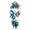
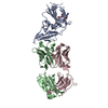
Mass: 23191.023 Da / Num. of mol.: 2 / Fragment: residues 481-650
Source method: isolated from a genetically manipulated source
Source: (gene. exp.) TGEV virulent Purdue (virus) / Strain: SC11 / Gene: S / Plasmid: pBJ5-GS / Cell line (production host): CHO Lec 3.2.8.1 / Production host: Cricetulus griseus (Chinese hamster) / References: UniProt: Q0PKZ5

-
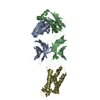
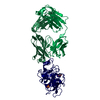
Antibody , 2 types, 4 molecules ACBD

#1: Antibody monoclonal antibody 1AF10, heavy chain


Mass: 23643.396 Da / Num. of mol.: 2 / Fragment: Fab
Source method: isolated from a genetically manipulated source
Source: (gene. exp.) Mus musculus (house mouse) / Description: Fab fragment prepared by papain digestion / Cell: hybridoma
#2: Antibody monoclonal antibody 1AF10, light chain


Mass: 23484.758 Da / Num. of mol.: 2 / Fragment: Fab
Source method: isolated from a genetically manipulated source
Source: (gene. exp.) Mus musculus (house mouse) / Description: Fab fragment prepared by papain digestion / Cell: hybridoma

-
Sugars , 2 types, 5 molecules

#4: Polysaccharide
2-acetamido-2-deoxy-beta-D-glucopyranose-(1-4)-2-acetamido-2-deoxy-beta-D-glucopyranose


Type: oligosaccharide / Mass: 424.401 Da / Num. of mol.: 4
Source method: isolated from a genetically manipulated source
DescriptorTypeProgram
DGlcpNAcb1-4DGlcpNAcb1-Glycam Condensed SequenceGMML 1.0
WURCS=2.0/1,2,1/[a2122h-1b_1-5_2*NCC/3=O]/1-1/a4-b1WURCSPDB2Glycan 1.1.0
[]{[(4+1)][b-D-GlcpNAc]{[(4+1)][b-D-GlcpNAc]{}}}LINUCSPDB-CARE
#6: Sugar ChemComp-NAG / 2-acetamido-2-deoxy-beta-D-glucopyranose / N-Acetylglucosamine


Type: D-saccharide, beta linking / Mass: 221.208 Da / Num. of mol.: 1
Source method: isolated from a genetically manipulated source
Formula: C8H15NO6
IdentifierTypeProgram
DGlcpNAcbCONDENSED IUPAC CARBOHYDRATE SYMBOLGMML 1.0
N-acetyl-b-D-glucopyranosamineCOMMON NAMEGMML 1.0
b-D-GlcpNAcIUPAC CARBOHYDRATE SYMBOLPDB-CARE 1.0
GlcNAcSNFG CARBOHYDRATE SYMBOLGMML 1.0

-
Non-polymers , 2 types, 4 molecules

#5: Chemical ChemComp-ACY / ACETIC ACID / Acetic acid


Mass: 60.052 Da / Num. of mol.: 2 / Source method: obtained synthetically / Formula: C2H4O2
#7: Water ChemComp-HOH / water / Water


Mass: 18.015 Da / Num. of mol.: 2 / Source method: isolated from a natural source / Formula: H2O

-
Experimental details

-
Experiment

ExperimentMethod: X-RAY DIFFRACTION / Number of used crystals: 1

-
Sample preparation

CrystalDensity Matthews: 3.22 Å3/Da / Density % sol: 61.77 %
Crystal growTemperature: 294 K / Method: vapor diffusion, hanging drop / pH: 8.5
Details: 16% PEG4000, 0.2 M sodium acetate, 0.1 M 1,2,3-octanetriol isomer T, 0.1 M Tris, pH 8.5, VAPOR DIFFUSION, HANGING DROP, temperature 294K

-
Data collection

DiffractionMean temperature: 200 K
Diffraction sourceSource: SYNCHROTRON / Site: ESRF / Beamline: ID29 / Wavelength: 0.97934 Å
DetectorType: ADSC QUANTUM 315r / Detector: CCD / Date: Feb 27, 2007
RadiationMonochromator: Si(111) / Protocol: SINGLE WAVELENGTH / Monochromatic (M) / Laue (L): M / Scattering type: x-ray
Radiation wavelengthWavelength: 0.97934 Å / Relative weight: 1
ReflectionResolution: 3→25 Å / Num. all: 35787 / Num. obs: 33913 / % possible obs: 94.7 % / Observed criterion σ(F): 1 / Observed criterion σ(I): 1 / Redundancy: 5.6 % / Biso Wilson estimate: 82 Å2 / Rmerge(I) obs: 0.074 / Rsym value: 0.074 / Net I/σ(I): 8.4
Reflection shellResolution: 3→3.16 Å / Redundancy: 5.2 % / Rmerge(I) obs: 0.354 / Mean I/σ(I) obs: 2 / Num. unique all: 4997 / Rsym value: 0.354 / % possible all: 95.3

-
Processing

Software
NameVersionClassification
DNAdata collection
PHASERphasing
PHENIX(phenix.refine: 1.6.4_486)refinement
XDSdata reduction
SCALAdata scaling
RefinementMethod to determine structure: MOLECULAR REPLACEMENT / Resolution: 3→25 Å / SU ML: 0.4 / σ(F): 1 / Phase error: 27.24 / Stereochemistry target values: ML
RfactorNum. reflection% reflectionSelection details
Rfree0.2507 1704 5.05 %RANDOM
Rwork0.216 ---
obs0.2178 33913 94.7 %-
all-35787 --
Solvent computationShrinkage radii: 0.9 Å / VDW probe radii: 1.11 Å / Solvent model: FLAT BULK SOLVENT MODEL / Bsol: 25.586 Å2 / ksol: 0.264 e/Å3
Displacement parameters
Baniso -1Baniso -2Baniso -3
1--8.9591 Å2-0 Å215.3985 Å2
2--14.9343 Å20 Å2
3----5.9752 Å2
Refinement stepCycle: LAST / Resolution: 3→25 Å
ProteinNucleic acidLigandSolventTotal
Num. atoms8862 0 134 2 8998
Refine LS restraints
Refine-IDTypeDev idealNumber
X-RAY DIFFRACTIONf_bond_d0.0059226
X-RAY DIFFRACTIONf_angle_d1.20712549
X-RAY DIFFRACTIONf_dihedral_angle_d25.5985612
X-RAY DIFFRACTIONf_chiral_restr0.0811441
X-RAY DIFFRACTIONf_plane_restr0.0061582
LS refinement shell
Resolution (Å)Rfactor RfreeNum. reflection RfreeRfactor RworkNum. reflection RworkRefine-ID% reflection obs (%)
3-3.04680.4021510.35652577X-RAY DIFFRACTION85
3.0468-3.09670.3921390.33782558X-RAY DIFFRACTION85
3.0967-3.150.36791250.32662597X-RAY DIFFRACTION85
3.15-3.20710.3461430.3092565X-RAY DIFFRACTION85
3.2071-3.26870.30731550.29392545X-RAY DIFFRACTION86
3.2687-3.33530.35151330.29072587X-RAY DIFFRACTION84
3.3353-3.40760.24861430.26632520X-RAY DIFFRACTION85
3.4076-3.48670.30491650.26232533X-RAY DIFFRACTION85
3.4867-3.57360.27951220.26992583X-RAY DIFFRACTION84
3.5736-3.66990.32231140.26542560X-RAY DIFFRACTION84
3.6699-3.77750.34131280.25782577X-RAY DIFFRACTION84
3.7775-3.8990.24971210.23752538X-RAY DIFFRACTION84
3.899-4.03780.251380.21662538X-RAY DIFFRACTION85
4.0378-4.19870.24851300.19852540X-RAY DIFFRACTION83
4.1987-4.38880.22431370.18082517X-RAY DIFFRACTION83
4.3888-4.61870.2211480.16762527X-RAY DIFFRACTION83
4.6187-4.9060.17391210.15522521X-RAY DIFFRACTION83
4.906-5.28130.21451520.16762889X-RAY DIFFRACTION95
5.2813-5.80650.20221510.17613012X-RAY DIFFRACTION100
5.8065-6.63240.28481480.21093012X-RAY DIFFRACTION100
6.6324-8.30270.22991680.19173020X-RAY DIFFRACTION100
8.3027-24.64270.17861620.1852880X-RAY DIFFRACTION95
Refinement TLS params.

Method: refined / Refine-ID: X-RAY DIFFRACTION

IDL112)L122)L132)L222)L232)L332)S11 (Å °)S12 (Å °)S13 (Å °)S21 (Å °)S22 (Å °)S23 (Å °)S31 (Å °)S32 (Å °)S33 (Å °)T112)T122)T132)T222)T232)T332)Origin x (Å)Origin y (Å)Origin z (Å)
11.3748-0.3961-0.83041.16740.25281.09980.01010.01830.07390.01630.1054-0.0672-0.19290.123400.2288-0.0504-0.10710.2080.0450.1789-93.444318.5847102.7875
21.44371.0591-0.56960.9689-0.00750.4458-0.02920.47160.0388-0.1233-0.15740.21180.217-0.44610.00580.3557-0.1239-0.07380.3675-0.0870.3232-107.61261.951398.1823
30.9779-0.11110.00081.3940.36921.24140.1708-0.4690.47-0.03190.1927-0.059-0.32690.20870.00050.4689-0.09470.1070.2477-0.15810.3985-10.314813.408632.7423
41.2268-1.64720.0551.47740.45450.8882-0.2011-0.2259-0.22860.17560.16760.22470.3126-0.2440.01450.3086-0.1196-0.01410.247-0.03870.331-15.2107-7.265425.7649
50.54380.139-0.68740.79510.38130.73830.49720.25210.48090.6044-0.20910.9901-0.5163-0.6540.0270.4855-0.07830.2630.5153-0.01920.8123-125.704628.1846118.834
61.12550.0814-1.93051.0908-0.61232.43590.33240.81830.288-0.37350.04911.2398-0.2263-1.02031.10690.41080.1212-0.1660.92640.17531.0381-134.414425.7389105.4055
71.03970.50611.10050.96660.06312.04480.23880.04540.3756-0.26280.0908-0.4963-0.01750.7601-0.04030.5088-0.22190.21110.6325-0.18030.579321.22030.600215.6874
80.9747-0.6308-0.27610.2969-0.07140.80120.146-0.70090.2064-0.04290.1452-0.0092-0.03310.58220.00890.3582-0.0567-0.04040.5814-0.160.207120.4281-12.770423.9432
91.497-1.4135-0.9186-0.6252-0.04990.9096-0.10520.75920.0372-0.0914-0.2199-0.05370.23-0.3286-0.00010.5259-0.05910.17750.4330.01590.4864-71.2531-0.255374.8216
102.2910.0329-1.61610.755-0.06621.85490.28440.74060.36110.14590.0628-0.0164-0.4247-0.37570.05260.39640.06530.03340.7461-0.01730.2267-45.41372.350750.2086
Refinement TLS group
IDRefine-IDRefine TLS-IDSelection details
1X-RAY DIFFRACTION1chain A and resid 1:122
2X-RAY DIFFRACTION2chain B and resid 1:108
3X-RAY DIFFRACTION3chain C and resid 1:122
4X-RAY DIFFRACTION4chain D and resid 1:108
5X-RAY DIFFRACTION5chain A and resid 123:219
6X-RAY DIFFRACTION6chain B and resid 109:213
7X-RAY DIFFRACTION7chain C and resid 123:220
8X-RAY DIFFRACTION8chain D and resid 109:211
9X-RAY DIFFRACTION9chain E
10X-RAY DIFFRACTION10chain F

+
About Yorodumi

-
News

-
Feb 9, 2022. New format data for meta-information of EMDB entries

New format data for meta-information of EMDB entries

  • Version 3 of the EMDB header file is now the official format.
  • The previous official version 1.9 will be removed from the archive.

Related info.:EMDB header

External links:wwPDB to switch to version 3 of the EMDB data model

-
Aug 12, 2020. Covid-19 info

Covid-19 info

URL: https://pdbj.org/emnavi/covid19.php

New page: Covid-19 featured information page in EM Navigator.

Related info.:Covid-19 info / Mar 5, 2020. Novel coronavirus structure data

+
Mar 5, 2020. Novel coronavirus structure data

Novel coronavirus structure data

Related info.:Yorodumi Speices / Aug 12, 2020. Covid-19 info

External links:COVID-19 featured content - PDBj / Molecule of the Month (242):Coronavirus Proteases

+
Jan 31, 2019. EMDB accession codes are about to change! (news from PDBe EMDB page)

EMDB accession codes are about to change! (news from PDBe EMDB page)

  • The allocation of 4 digits for EMDB accession codes will soon come to an end. Whilst these codes will remain in use, new EMDB accession codes will include an additional digit and will expand incrementally as the available range of codes is exhausted. The current 4-digit format prefixed with “EMD-” (i.e. EMD-XXXX) will advance to a 5-digit format (i.e. EMD-XXXXX), and so on. It is currently estimated that the 4-digit codes will be depleted around Spring 2019, at which point the 5-digit format will come into force.
  • The EM Navigator/Yorodumi systems omit the EMD- prefix.

Related info.:Q: What is EMD? / ID/Accession-code notation in Yorodumi/EM Navigator

External links:EMDB Accession Codes are Changing Soon! / Contact to PDBj

+
Jul 12, 2017. Major update of PDB

Major update of PDB

  • wwPDB released updated PDB data conforming to the new PDBx/mmCIF dictionary.
  • This is a major update changing the version number from 4 to 5, and with Remediation, in which all the entries are updated.
  • In this update, many items about electron microscopy experimental information are reorganized (e.g. em_software).
  • Now, EM Navigator and Yorodumi are based on the updated data.

External links:wwPDB Remediation / Enriched Model Files Conforming to OneDep Data Standards Now Available in the PDB FTP Archive

-
Yorodumi

Thousand views of thousand structures

  • Yorodumi is a browser for structure data from EMDB, PDB, SASBDB, etc.
  • This page is also the successor to EM Navigator detail page, and also detail information page/front-end page for Omokage search.
  • The word "yorodu" (or yorozu) is an old Japanese word meaning "ten thousand". "mi" (miru) is to see.

Related info.:EMDB / PDB / SASBDB / Comparison of 3 databanks / Yorodumi Search / Aug 31, 2016. New EM Navigator & Yorodumi / Yorodumi Papers / Jmol/JSmol / Function and homology information / Changes in new EM Navigator and Yorodumi

Read more