 Movie
Movie Controller

[English] 日本語
 Yorodumi
- PDB-4f22: Kainate bound to the K660A mutant of the ligand binding domain of... -
+ Open data

- Basic information
| Entry | Database: PDB / ID: 4f22 | ||||||
|---|---|---|---|---|---|---|---|
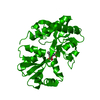
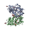
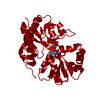
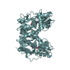
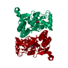
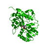
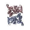
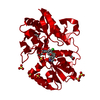
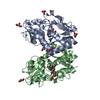
| Title | Kainate bound to the K660A mutant of the ligand binding domain of GluA3 | ||||||
 Components Components | Glutamate receptor 3 | ||||||
 Keywords Keywords | TRANSPORT PROTEIN/AGONIST / glutamate receptor / GluA3 / GluR3 / AMPA receptor / S1S2 / LBD / neurotransmitter receptor / kainate / TRANSPORT PROTEIN / TRANSPORT PROTEIN-AGONIST complex | ||||||
| Function / homology |  Function and homology information Function and homology informationchemical synaptic transmission, postsynaptic / Trafficking of AMPA receptors / Synaptic adhesion-like molecules / parallel fiber to Purkinje cell synapse / Activation of AMPA receptors / response to lithium ion / AMPA glutamate receptor activity / Trafficking of GluR2-containing AMPA receptors / AMPA glutamate receptor complex / ionotropic glutamate receptor complex ...chemical synaptic transmission, postsynaptic / Trafficking of AMPA receptors / Synaptic adhesion-like molecules / parallel fiber to Purkinje cell synapse / Activation of AMPA receptors / response to lithium ion / AMPA glutamate receptor activity / Trafficking of GluR2-containing AMPA receptors / AMPA glutamate receptor complex / ionotropic glutamate receptor complex / asymmetric synapse / regulation of receptor recycling / Unblocking of NMDA receptors, glutamate binding and activation / regulation of postsynaptic membrane potential / glutamate-gated receptor activity / synaptic cleft / presynaptic active zone membrane / response to fungicide / ligand-gated monoatomic ion channel activity involved in regulation of presynaptic membrane potential / monoatomic ion transmembrane transport / transmitter-gated monoatomic ion channel activity involved in regulation of postsynaptic membrane potential / dendritic shaft / synaptic transmission, glutamatergic / postsynaptic density membrane / modulation of chemical synaptic transmission / terminal bouton / presynaptic membrane / amyloid-beta binding / perikaryon / chemical synaptic transmission / postsynaptic membrane / dendritic spine / postsynaptic density / neuronal cell body / dendrite / glutamatergic synapse / protein-containing complex / membrane / plasma membraneSimilarity search - Function | ||||||
| Biological species |  Rattus norvegicus (Norway rat) Rattus norvegicus (Norway rat) | ||||||
| Method | X-RAY DIFFRACTION / SYNCHROTRON / MOLECULAR REPLACEMENT / Resolution: 2.06 Å | ||||||
 Authors Authors | Ahmed, A.H. / Oswald, R.E. | ||||||
 Citation Citation | Journal: Biochemistry / Year: 2012 Title: The loss of an electrostatic contact unique to AMPA receptor ligand binding domain 2 slows channel activation. Authors: Holley, S.M. / Ahmed, A.H. / Srinivasan, J. / Murthy, S.E. / Weiland, G.A. / Oswald, R.E. / Nowak, L.M. | ||||||
| History |
|
- Structure visualization
| Structure viewer | Molecule: MolmilJmol/JSmol |
|---|
- Downloads & links
-Download
| PDBx/mmCIF format | 4f22.cif.gz | 71 KB | Display | PDBx/mmCIF format |
|---|---|---|---|---|
| PDB format | pdb4f22.ent.gz | 50.6 KB | Display | PDB format |
| PDBx/mmJSON format | 4f22.json.gz | Tree view | PDBx/mmJSON format | |
| Others |  Other downloads Other downloads |
-Validation report
| Arichive directory | https://data.pdbj.org/pub/pdb/validation_reports/f2/4f22ftp://data.pdbj.org/pub/pdb/validation_reports/f2/4f22 | HTTPS FTP |
|---|
-Related structure data
| Related structure data |  4f1yC  4f29C  4f2oC  4f2qC  4f31C  4f39C 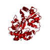 4f3bC  4f3gC  3dlnS C: citing same article ( S: Starting model for refinement |
|---|---|
| Similar structure data |
-Links
 PDBj
PDBj


- Assembly
| Deposited unit | 
| ||||||||
|---|---|---|---|---|---|---|---|---|---|
| 1 | 
| ||||||||
| Unit cell |
|
-Components
| #1: Protein | / GluR-3 / AMPA-selective glutamate receptor 3 / GluR-C / GluR-K3 / Glutamate receptor ionotropic / ...GluR-3 / AMPA-selective glutamate receptor 3 / GluR-C / GluR-K3 / Glutamate receptor ionotropic / AMPA 3 / GluA3 Mass: 28898.316 Da / Num. of mol.: 1 / Mutation: K660A Source method: isolated from a genetically manipulated source Source: (gene. exp.) Rattus norvegicus (Norway rat) / Gene: Glur3, Gria3, Gria3; GluA3 / Plasmid: pET-22b(+) / Production host:  Escherichia coli (E. coli) / Strain (production host): Origami B (DE3) / References: UniProt: P19492 Escherichia coli (E. coli) / Strain (production host): Origami B (DE3) / References: UniProt: P19492 |
|---|---|
| #2: Chemical | ChemComp-KAI / Kainic acid  Mass: 213.230 Da / Num. of mol.: 1 / Source method: obtained synthetically / Formula: C10H15NO4 / Comment: neurotransmitter, agonist*YM Mass: 213.230 Da / Num. of mol.: 1 / Source method: obtained synthetically / Formula: C10H15NO4 / Comment: neurotransmitter, agonist*YM |
| #3: Chemical | ChemComp-ZN /   Mass: 65.409 Da / Num. of mol.: 1 / Source method: obtained synthetically / Formula: Zn Mass: 65.409 Da / Num. of mol.: 1 / Source method: obtained synthetically / Formula: Zn |
| #4: Water | ChemComp-HOH / Water Mass: 18.015 Da / Num. of mol.: 235 / Source method: isolated from a natural source / Formula: H2O Mass: 18.015 Da / Num. of mol.: 235 / Source method: isolated from a natural source / Formula: H2O |
| Sequence details | RESIDUES 232,233, 242, AND 246 CORRESPOND |
-Experimental details
-Experiment
| Experiment | Method: X-RAY DIFFRACTION / Number of used crystals: 1 |
|---|
- Sample preparation
| Crystal | Density Matthews: 2.76 Å3/Da / Density % sol: 55.47 % |
|---|---|
| Crystal grow | Temperature: 277 K / Method: vapor diffusion, hanging drop / pH: 6.5 Details: 14-15% PEG 8K, 0.1 M sodium cacodylate, 0.1-0.15 M zinc acetate, 0.25 M ammonium sulfate, pH 6.5, VAPOR DIFFUSION, HANGING DROP, temperature 277K |
-Data collection
| Diffraction | Mean temperature: 100 K |
|---|---|
| Diffraction source | Source: SYNCHROTRON / Site: CHESS  / Beamline: A1 / Wavelength: 0.977 Å / Beamline: A1 / Wavelength: 0.977 Å |
| Detector | Type: ADSC QUANTUM 210 / Detector: CCD / Date: Dec 5, 2010 |
| Radiation | Monochromator: Rh coated Si / Protocol: SINGLE WAVELENGTH / Monochromatic (M) / Laue (L): M / Scattering type: x-ray |
| Radiation wavelength | Wavelength: 0.977 Å / Relative weight: 1 |
| Reflection | Resolution: 2.06→50 Å / % possible obs: 99.7 % / Observed criterion σ(F): 0 / Observed criterion σ(I): -3 / Redundancy: 5.1 % / Rmerge(I) obs: 0.157 / Rsym value: 0.157 / Net I/σ(I): 11.112 |
| Reflection shell | Resolution: 2.08→2.12 Å / Redundancy: 4.5 % / Rmerge(I) obs: 0.312 / Mean I/σ(I) obs: 2.261 / Rsym value: 0.312 / % possible all: 100 |
- Processing
| Software |
| ||||||||||||||||||||||||||||||||||||||||||||||||||||||||||||||||||||||||||||||||||||||||||||||||||||||||||||||||||||||||||||||||||||||||||||||||||||||||||||||||||||||||||
|---|---|---|---|---|---|---|---|---|---|---|---|---|---|---|---|---|---|---|---|---|---|---|---|---|---|---|---|---|---|---|---|---|---|---|---|---|---|---|---|---|---|---|---|---|---|---|---|---|---|---|---|---|---|---|---|---|---|---|---|---|---|---|---|---|---|---|---|---|---|---|---|---|---|---|---|---|---|---|---|---|---|---|---|---|---|---|---|---|---|---|---|---|---|---|---|---|---|---|---|---|---|---|---|---|---|---|---|---|---|---|---|---|---|---|---|---|---|---|---|---|---|---|---|---|---|---|---|---|---|---|---|---|---|---|---|---|---|---|---|---|---|---|---|---|---|---|---|---|---|---|---|---|---|---|---|---|---|---|---|---|---|---|---|---|---|---|---|---|---|---|---|
| Refinement | Method to determine structure: MOLECULAR REPLACEMENT Starting model: 3DLN Resolution: 2.06→33.15 Å / Cor.coef. Fo:Fc: 0.953 / Cor.coef. Fo:Fc free: 0.923 / SU B: 4.508 / SU ML: 0.124 / Isotropic thermal model: Isotropic / Cross valid method: THROUGHOUT / ESU R: 0.189 / ESU R Free: 0.181 / Stereochemistry target values: MAXIMUM LIKELIHOOD / Details: HYDROGENS HAVE BEEN ADDED IN THE RIDING POSITIONS
| ||||||||||||||||||||||||||||||||||||||||||||||||||||||||||||||||||||||||||||||||||||||||||||||||||||||||||||||||||||||||||||||||||||||||||||||||||||||||||||||||||||||||||
| Solvent computation | Ion probe radii: 0.8 Å / Shrinkage radii: 0.8 Å / VDW probe radii: 1.4 Å / Solvent model: MASK | ||||||||||||||||||||||||||||||||||||||||||||||||||||||||||||||||||||||||||||||||||||||||||||||||||||||||||||||||||||||||||||||||||||||||||||||||||||||||||||||||||||||||||
| Displacement parameters | Biso mean: 22.342 Å2
| ||||||||||||||||||||||||||||||||||||||||||||||||||||||||||||||||||||||||||||||||||||||||||||||||||||||||||||||||||||||||||||||||||||||||||||||||||||||||||||||||||||||||||
| Refinement step | Cycle: LAST / Resolution: 2.06→33.15 Å
| ||||||||||||||||||||||||||||||||||||||||||||||||||||||||||||||||||||||||||||||||||||||||||||||||||||||||||||||||||||||||||||||||||||||||||||||||||||||||||||||||||||||||||
| Refine LS restraints |
| ||||||||||||||||||||||||||||||||||||||||||||||||||||||||||||||||||||||||||||||||||||||||||||||||||||||||||||||||||||||||||||||||||||||||||||||||||||||||||||||||||||||||||
| LS refinement shell | Resolution: 2.064→2.118 Å / Total num. of bins used: 20
|
