 Movie
Movie Controller

[English] 日本語
 Yorodumi
- PDB-4f1h: Crystal structure of TDP2 from Danio rerio complexed with a singl... -
+ Open data

- Basic information
| Entry | Database: PDB / ID: 4f1h | ||||||
|---|---|---|---|---|---|---|---|
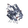
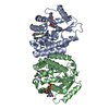
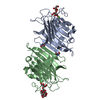
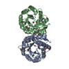
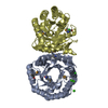
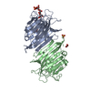
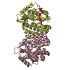
| Title | Crystal structure of TDP2 from Danio rerio complexed with a single strand DNA | ||||||
 Components Components |
| ||||||
 Keywords Keywords | Hydrolase/DNA / 5'-tyrosyl DNA phosphodiesterase / Hydrolase-DNA complex | ||||||
| Function / homology |  Function and homology information Function and homology informationtyrosyl-DNA phosphodiesterase activity / : / convergent extension involved in gastrulation / 5'-tyrosyl-DNA phosphodiesterase activity /  nuclease activity / Hydrolases; Acting on ester bonds; Phosphoric-diester hydrolases / determination of left/right symmetry / gastrulation / negative regulation of transforming growth factor beta receptor signaling pathway / PML body ...tyrosyl-DNA phosphodiesterase activity / : / convergent extension involved in gastrulation / 5'-tyrosyl-DNA phosphodiesterase activity / nuclease activity / Hydrolases; Acting on ester bonds; Phosphoric-diester hydrolases / determination of left/right symmetry / gastrulation / negative regulation of transforming growth factor beta receptor signaling pathway / PML body / double-strand break repair / single-stranded DNA binding / manganese ion binding / magnesium ion binding / DNA binding / cytoplasm nuclease activity / Hydrolases; Acting on ester bonds; Phosphoric-diester hydrolases / determination of left/right symmetry / gastrulation / negative regulation of transforming growth factor beta receptor signaling pathway / PML body ...tyrosyl-DNA phosphodiesterase activity / : / convergent extension involved in gastrulation / 5'-tyrosyl-DNA phosphodiesterase activity / nuclease activity / Hydrolases; Acting on ester bonds; Phosphoric-diester hydrolases / determination of left/right symmetry / gastrulation / negative regulation of transforming growth factor beta receptor signaling pathway / PML body / double-strand break repair / single-stranded DNA binding / manganese ion binding / magnesium ion binding / DNA binding / cytoplasmSimilarity search - Function | ||||||
| Biological species |  Danio rerio (zebrafish) Danio rerio (zebrafish) | ||||||
| Method | X-RAY DIFFRACTION / SYNCHROTRON / MOLECULAR REPLACEMENT / Resolution: 1.662 Å | ||||||
 Authors Authors | Shi, K. / Kurahashi, K. / Aihara, H. | ||||||
 Citation Citation | Journal: Nat.Struct.Mol.Biol. / Year: 2012 Title: Structural basis for recognition of 5'-phosphotyrosine adducts by Tdp2. Authors: Shi, K. / Kurahashi, K. / Gao, R. / Tsutakawa, S.E. / Tainer, J.A. / Pommier, Y. / Aihara, H. | ||||||
| History |
|
- Structure visualization
| Structure viewer | Molecule: MolmilJmol/JSmol |
|---|
- Downloads & links
-Download
| PDBx/mmCIF format | 4f1h.cif.gz | 245.8 KB | Display | PDBx/mmCIF format |
|---|---|---|---|---|
| PDB format | pdb4f1h.ent.gz | 194.1 KB | Display | PDB format |
| PDBx/mmJSON format | 4f1h.json.gz | Tree view | PDBx/mmJSON format | |
| Others |  Other downloads Other downloads |
-Validation report
| Arichive directory | https://data.pdbj.org/pub/pdb/validation_reports/f1/4f1hftp://data.pdbj.org/pub/pdb/validation_reports/f1/4f1h | HTTPS FTP |
|---|
-Related structure data


-Links
 PDBj
PDBj


- Assembly
| Deposited unit | 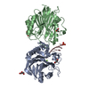
| ||||||||
|---|---|---|---|---|---|---|---|---|---|
| 1 |
| ||||||||
| Unit cell |
|
-Components
-DNA chain , 1 types, 1 molecules C
| #1: DNA chain | Mass: 1520.036 Da / Num. of mol.: 1 / Source method: obtained synthetically |
|---|
-Tyrosyl-DNA phosphodiesterase ... , 2 types, 2 molecules AB
| #2: Protein | Mass: 28484.727 Da / Num. of mol.: 1 Source method: isolated from a genetically manipulated source Source: (gene. exp.) Danio rerio (zebrafish)Gene: tdp2, ttrap, ttrapl, si:ch211-81e5.5, si:dkey-218n20.5 Production host:  Escherichia coli (E. coli) Escherichia coli (E. coli)References: UniProt: Q5XJA0, Hydrolases; Acting on ester bonds; Phosphoric-diester hydrolases |
|---|---|
| #3: Protein | Mass: 28613.836 Da / Num. of mol.: 1 Source method: isolated from a genetically manipulated source Source: (gene. exp.) Danio rerio (zebrafish)Gene: tdp2, ttrap, ttrapl, si:ch211-81e5.5, si:dkey-218n20.5 Production host: Escherichia coli (E. coli)References: UniProt: Q5XJA0, Hydrolases; Acting on ester bonds; Phosphoric-diester hydrolases |
-Non-polymers , 4 types, 592 molecules 


| #4: Chemical | ChemComp-PO4 / Phosphate Mass: 94.971 Da / Num. of mol.: 1 / Source method: obtained synthetically / Formula: PO4 Mass: 94.971 Da / Num. of mol.: 1 / Source method: obtained synthetically / Formula: PO4 | ||||
|---|---|---|---|---|---|
| #5: Chemical | ChemComp-GOL / Glycerol Mass: 92.094 Da / Num. of mol.: 4 / Source method: obtained synthetically / Formula: C3H8O3 Mass: 92.094 Da / Num. of mol.: 4 / Source method: obtained synthetically / Formula: C3H8O3#6: Chemical | ChemComp-MG / |  Mass: 24.305 Da / Num. of mol.: 1 / Source method: obtained synthetically / Formula: Mg Mass: 24.305 Da / Num. of mol.: 1 / Source method: obtained synthetically / Formula: Mg#7: Water | ChemComp-HOH / | WaterMass: 18.015 Da / Num. of mol.: 586 / Source method: isolated from a natural source / Formula: H2O |
-Details
| Sequence details | A LARGE PORTION OF THE N-TERMINAL WAS NOT OBSERVED IN THE ELECTRON DENSITY |
|---|
-Experimental details
-Experiment
| Experiment | Method: X-RAY DIFFRACTION / Number of used crystals: 1 |
|---|
- Sample preparation
| Crystal | Density Matthews: 2.46 Å3/Da / Density % sol: 50.02 % |
|---|---|
| Crystal grow | Temperature: 293 K / pH: 7 Details: PEG and sodium tartrate, pH 7, VAPOR DIFFUSION, SITTING DROP, temperature 293K |
-Data collection
| Diffraction | Mean temperature: 96 K |
|---|---|
| Diffraction source | Source: SYNCHROTRON / Site: APS  / Beamline: 24-ID-C / Wavelength: 0.979 / Beamline: 24-ID-C / Wavelength: 0.979 |
| Detector | Type: ADSC QUANTUM 315 / Detector: CCD / Date: Dec 9, 2011 |
| Radiation | Monochromator: SI 111 CHANNEL / Protocol: SINGLE WAVELENGTH / Monochromatic (M) / Laue (L): M / Scattering type: x-ray |
| Radiation wavelength | Wavelength: 0.979 Å / Relative weight: 1 |
| Reflection | Resolution: 1.66→50 Å / Num. obs: 67804 / % possible obs: 99.4 % / Observed criterion σ(I): 0 |
- Processing
| Software |
| |||||||||||||||||||||||||||||||||||||||||||||||||||||||||||||||||||||||||||||||||||||||||||||||||||||||||
|---|---|---|---|---|---|---|---|---|---|---|---|---|---|---|---|---|---|---|---|---|---|---|---|---|---|---|---|---|---|---|---|---|---|---|---|---|---|---|---|---|---|---|---|---|---|---|---|---|---|---|---|---|---|---|---|---|---|---|---|---|---|---|---|---|---|---|---|---|---|---|---|---|---|---|---|---|---|---|---|---|---|---|---|---|---|---|---|---|---|---|---|---|---|---|---|---|---|---|---|---|---|---|---|---|---|---|
| Refinement | Method to determine structure: MOLECULAR REPLACEMENT / Resolution: 1.662→45.871 Å / SU ML: 0.2 / σ(F): 1.35 / Phase error: 17.52 / Stereochemistry target values: ML
| |||||||||||||||||||||||||||||||||||||||||||||||||||||||||||||||||||||||||||||||||||||||||||||||||||||||||
| Solvent computation | Shrinkage radii: 0.73 Å / VDW probe radii: 1 Å / Solvent model: FLAT BULK SOLVENT MODEL / Bsol: 44.239 Å2 / ksol: 0.353 e/Å3 | |||||||||||||||||||||||||||||||||||||||||||||||||||||||||||||||||||||||||||||||||||||||||||||||||||||||||
| Displacement parameters |
| |||||||||||||||||||||||||||||||||||||||||||||||||||||||||||||||||||||||||||||||||||||||||||||||||||||||||
| Refinement step | Cycle: LAST / Resolution: 1.662→45.871 Å
| |||||||||||||||||||||||||||||||||||||||||||||||||||||||||||||||||||||||||||||||||||||||||||||||||||||||||
| Refine LS restraints |
| |||||||||||||||||||||||||||||||||||||||||||||||||||||||||||||||||||||||||||||||||||||||||||||||||||||||||
| LS refinement shell |
| |||||||||||||||||||||||||||||||||||||||||||||||||||||||||||||||||||||||||||||||||||||||||||||||||||||||||
| Refinement TLS params. | Method: refined / Origin x: 16.7302 Å / Origin y: -2.4536 Å / Origin z: -1.7162 Å
| |||||||||||||||||||||||||||||||||||||||||||||||||||||||||||||||||||||||||||||||||||||||||||||||||||||||||
| Refinement TLS group | Selection details: all |
