 Movie
Movie Controller

[English] 日本語
 Yorodumi
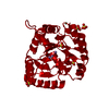
- PDB-3lm7: Crystal Structure of DUF1341 representative, from Yersinia entero... -
+ Open data

- Basic information
| Entry | Database: PDB / ID: 3lm7 | ||||||
|---|---|---|---|---|---|---|---|
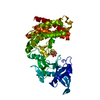
| Title | Crystal Structure of DUF1341 representative, from Yersinia enterocolitica subsp. enterocolitica 8081 | ||||||
 Components Components | putative 4-Hydroxy-2-oxoglutarate aldolase / 2-dehydro-3-deoxyphosphogluconate aldolase | ||||||
 Keywords Keywords |  LYASE / putative 4-Hydroxy-oxoglutarate aldolase / 2-dehydro-3-deoxyphosphogluconate aldolase / Structural Genomics / PSI-2 / Protein Structure Initiative / Midwest Center for Structural Genomics / MCSG LYASE / putative 4-Hydroxy-oxoglutarate aldolase / 2-dehydro-3-deoxyphosphogluconate aldolase / Structural Genomics / PSI-2 / Protein Structure Initiative / Midwest Center for Structural Genomics / MCSG | ||||||
| Function / homology |  Function and homology information Function and homology information | ||||||
| Biological species |  Yersinia enterocolitica (bacteria) Yersinia enterocolitica (bacteria) | ||||||
| Method | X-RAY DIFFRACTION / SYNCHROTRON / MAD / Resolution: 1.9 Å | ||||||
 Authors Authors | Joachimiak, A. / Duke, N.E.C. / Feldmann, B. / Wu, R. / Midwest Center for Structural Genomics (MCSG) | ||||||
 Citation Citation | Journal: To be Published Title: Crystal Structure of DUF1341 representative, from Yersinia enterocolitica subsp. enterocolitica 8081 Authors: Joachimiak, A. / Duke, N.E.C. / Feldmann, B. / Wu, R. | ||||||
| History |
|
- Structure visualization
| Structure viewer | Molecule: MolmilJmol/JSmol |
|---|
- Downloads & links
-Download
| PDBx/mmCIF format | 3lm7.cif.gz | 117.1 KB | Display | PDBx/mmCIF format |
|---|---|---|---|---|
| PDB format | pdb3lm7.ent.gz | 96.4 KB | Display | PDB format |
| PDBx/mmJSON format | 3lm7.json.gz | Tree view | PDBx/mmJSON format | |
| Others |  Other downloads Other downloads |
-Validation report
| Arichive directory | https://data.pdbj.org/pub/pdb/validation_reports/lm/3lm7ftp://data.pdbj.org/pub/pdb/validation_reports/lm/3lm7 | HTTPS FTP |
|---|
-Related structure data
| Similar structure data | |
|---|---|
| Other databases |


-Links
 PDBj
PDBj

- Assembly
| Deposited unit | 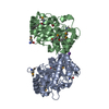
| ||||||||
|---|---|---|---|---|---|---|---|---|---|
| 1 |
| ||||||||
| 2 | 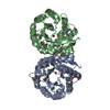
| ||||||||
| Unit cell |
| ||||||||
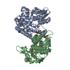
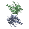
| Details | authors state that the biological unit is the dimer in asymmetric unit |
-Components
| #1: Protein | Mass: 26958.734 Da / Num. of mol.: 2 Source method: isolated from a genetically manipulated source Source: (gene. exp.) Yersinia enterocolitica (bacteria) / Strain: enterocolitica 8081 / Gene: YE3779 / Plasmid: pMCSG19b / Production host: Escherichia coli (E. coli) / Strain (production host): BL21(de3) / References: UniProt: A1JRF3#2: Chemical | Bromide  Mass: 79.904 Da / Num. of mol.: 2 / Source method: obtained synthetically / Formula: Br Mass: 79.904 Da / Num. of mol.: 2 / Source method: obtained synthetically / Formula: Br#3: Chemical | ChemComp-K / |   Mass: 39.098 Da / Num. of mol.: 1 / Source method: obtained synthetically / Formula: K Mass: 39.098 Da / Num. of mol.: 1 / Source method: obtained synthetically / Formula: K#4: Water | ChemComp-HOH / | Water Mass: 18.015 Da / Num. of mol.: 720 / Source method: isolated from a natural source / Formula: H2O Mass: 18.015 Da / Num. of mol.: 720 / Source method: isolated from a natural source / Formula: H2O |
|---|
-Experimental details
-Experiment
| Experiment | Method: X-RAY DIFFRACTION / Number of used crystals: 1 |
|---|
- Sample preparation
| Crystal | Density Matthews: 2.23 Å3/Da / Density % sol: 44.79 % |
|---|---|
| Crystal grow | Temperature: 289 K / Method: vapor diffusion, sitting drop / pH: 7.6 Details: 0.15M potassium bromide, 30% w/v polyethylene glycol monomethyl ether 2000 (Hampton Index #96), pH 7.60, VAPOR DIFFUSION, SITTING DROP, temperature 289.0K |
-Data collection
| Diffraction | Mean temperature: 100 K | |||||||||
|---|---|---|---|---|---|---|---|---|---|---|
| Diffraction source | Source: SYNCHROTRON / Site: APS  / Beamline: 19-BM / Wavelength: 0.97915, 0.97942 / Beamline: 19-BM / Wavelength: 0.97915, 0.97942 | |||||||||
| Detector | Type: ADSC QUANTUM 210r / Detector: CCD / Date: Nov 18, 2009 / Details: Si (111) double crystal monochromator | |||||||||
| Radiation | Monochromator: Si (111) double crystal monochromator / Protocol: MAD / Monochromatic (M) / Laue (L): M / Scattering type: x-ray | |||||||||
| Radiation wavelength |
| |||||||||
| Reflection | Resolution: 1.9→100 Å / Num. all: 755369 / Num. obs: 702493 / % possible obs: 93 % / Observed criterion σ(F): 2 / Redundancy: 9.6 % / Rmerge(I) obs: 0.065 | |||||||||
| Reflection shell | Resolution: 1.9→1.95 Å / Redundancy: 5.8 % / Rmerge(I) obs: 0.272 / Mean I/σ(I) obs: 6.4 / Rsym value: 0.248 / % possible all: 95 |
- Processing
| Software |
| |||||||||||||||||||||||||||||||||||||||||||||||||||||||||||||||||
|---|---|---|---|---|---|---|---|---|---|---|---|---|---|---|---|---|---|---|---|---|---|---|---|---|---|---|---|---|---|---|---|---|---|---|---|---|---|---|---|---|---|---|---|---|---|---|---|---|---|---|---|---|---|---|---|---|---|---|---|---|---|---|---|---|---|---|
| Refinement | Method to determine structure: MAD / Resolution: 1.9→79.56 Å / Cor.coef. Fo:Fc: 0.965 / Cor.coef. Fo:Fc free: 0.934 / SU B: 2.507 / SU ML: 0.075 / Cross valid method: THROUGHOUT / ESU R: 0.127 / ESU R Free: 0.13 / Stereochemistry target values: MAXIMUM LIKELIHOOD / Details: HYDROGENS HAVE BEEN ADDED IN THE RIDING POSITIONS
| |||||||||||||||||||||||||||||||||||||||||||||||||||||||||||||||||
| Solvent computation | Ion probe radii: 0.8 Å / Shrinkage radii: 0.8 Å / VDW probe radii: 1.4 Å / Solvent model: MASK | |||||||||||||||||||||||||||||||||||||||||||||||||||||||||||||||||
| Displacement parameters | Biso mean: 13.943 Å2
| |||||||||||||||||||||||||||||||||||||||||||||||||||||||||||||||||
| Refinement step | Cycle: LAST / Resolution: 1.9→79.56 Å
| |||||||||||||||||||||||||||||||||||||||||||||||||||||||||||||||||
| Refine LS restraints |
| |||||||||||||||||||||||||||||||||||||||||||||||||||||||||||||||||
| LS refinement shell | Resolution: 1.9→1.95 Å / Total num. of bins used: 20
|
