 Movie
Movie Controller

[English] 日本語
 Yorodumi
- PDB-4eza: Crystal structure of the atypical phosphoinositide (aPI) binding ... -
+ Open data

- Basic information
| Entry | Database: PDB / ID: 4eza | ||||||
|---|---|---|---|---|---|---|---|
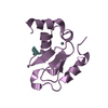
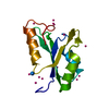
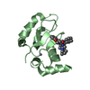
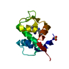
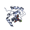
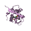
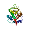
| Title | Crystal structure of the atypical phosphoinositide (aPI) binding domain of IQGAP2 | ||||||
 Components Components | Ras GTPase-activating-like protein IQGAP2 | ||||||
 Keywords Keywords |  SIGNALING PROTEIN / gap / structural genomics consortium / sgc SIGNALING PROTEIN / gap / structural genomics consortium / sgc | ||||||
| Function / homology |  Function and homology information Function and homology informationmitotic actomyosin contractile ring assembly actin filament organization / thrombin-activated receptor signaling pathway / Arp2/3 complex-mediated actin nucleation / Arp2/3 complex binding / GTPase inhibitor activity / microvillus / phosphatidylinositol-3,4,5-trisphosphate binding / CDC42 GTPase cycle / RHOG GTPase cycle / RHO GTPases activate IQGAPs ...mitotic actomyosin contractile ring assembly actin filament organization / thrombin-activated receptor signaling pathway / Arp2/3 complex-mediated actin nucleation / Arp2/3 complex binding / GTPase inhibitor activity / microvillus / phosphatidylinositol-3,4,5-trisphosphate binding / CDC42 GTPase cycle / RHOG GTPase cycle / RHO GTPases activate IQGAPs / RAC1 GTPase cycle / GTPase activator activity / secretory granule membrane / filopodium / regulation of actin cytoskeleton organization / small GTPase binding / actin filament binding / actin cytoskeleton / lamellipodium / actin binding / cell cortex / microtubule / calmodulin binding / Neutrophil degranulation / cell surface / signal transduction / extracellular exosome / plasma membrane / cytosol / cytoplasmSimilarity search - Function | ||||||
| Biological species |  Homo sapiens (human) Homo sapiens (human) | ||||||
| Method | X-RAY DIFFRACTION / SYNCHROTRON / Resolution: 1.5 Å | ||||||
 Authors Authors | Van Aalten, D.M.F. / Dixon, M.J. / Gray, A. / Schenning, M. / Agacan, M. / Leslie, N.R. / Downes, C.P. / Batty, I.H. / Nedyalkova, L. / Tempel, W. ...Van Aalten, D.M.F. / Dixon, M.J. / Gray, A. / Schenning, M. / Agacan, M. / Leslie, N.R. / Downes, C.P. / Batty, I.H. / Nedyalkova, L. / Tempel, W. / Tong, Y. / Zhong, N. / Crombet, L. / Arrowsmith, C.H. / Edwards, A.M. / Bountra, C. / Weigelt, J. / Bochkarev, A. / Park, H. / Structural Genomics Consortium (SGC) | ||||||
 Citation Citation | Journal: J.Biol.Chem. / Year: 2012 Title: IQGAP Proteins Reveal an Atypical Phosphoinositide (aPI) Binding Domain with a Pseudo C2 Domain Fold. Authors: Dixon, M.J. / Gray, A. / Schenning, M. / Agacan, M. / Tempel, W. / Tong, Y. / Nedyalkova, L. / Park, H.W. / Leslie, N.R. / van Aalten, D.M. / Downes, C.P. / Batty, I.H. | ||||||
| History |
|
- Structure visualization
| Structure viewer | Molecule: MolmilJmol/JSmol |
|---|
- Downloads & links
-Download
| PDBx/mmCIF format | 4eza.cif.gz | 93.2 KB | Display | PDBx/mmCIF format |
|---|---|---|---|---|
| PDB format | pdb4eza.ent.gz | 71.8 KB | Display | PDB format |
| PDBx/mmJSON format | 4eza.json.gz | Tree view | PDBx/mmJSON format | |
| Others |  Other downloads Other downloads |
-Validation report
| Arichive directory | https://data.pdbj.org/pub/pdb/validation_reports/ez/4ezaftp://data.pdbj.org/pub/pdb/validation_reports/ez/4eza | HTTPS FTP |
|---|
-Related structure data
| Similar structure data |
|---|


-Links
 PDBj
PDBj


- Assembly
| Deposited unit | 
| ||||||||
|---|---|---|---|---|---|---|---|---|---|
| 1 | 
| ||||||||
| 2 | 
| ||||||||
| Unit cell |
|
-Components
| #1: Protein | Mass: 13232.346 Da / Num. of mol.: 2 / Fragment: UNP residues 1476-1571 Source method: isolated from a genetically manipulated source Source: (gene. exp.) Homo sapiens (human) / Gene: IQGAP2 / Plasmid: pET28-mhl / Production host:  Escherichia coli (E. coli) / Strain (production host): BL21-V2R-pRARE2 / References: UniProt: Q13576 Escherichia coli (E. coli) / Strain (production host): BL21-V2R-pRARE2 / References: UniProt: Q13576#2: Water | ChemComp-HOH / | Water Mass: 18.015 Da / Num. of mol.: 156 / Source method: isolated from a natural source / Formula: H2O Mass: 18.015 Da / Num. of mol.: 156 / Source method: isolated from a natural source / Formula: H2O |
|---|
-Experimental details
-Experiment
| Experiment | Method: X-RAY DIFFRACTION / Number of used crystals: 1 |
|---|
- Sample preparation
| Crystal | Density Matthews: 1.87 Å3/Da / Density % sol: 34.11 % |
|---|---|
| Crystal grow | Temperature: 291 K / Method: vapor diffusion, sitting drop / pH: 6.5 Details: 30% PEG-MME, 0.2M ammonium sulfate, 0.1M MES, 1:100 (w/w) chymotrypsin, pH 6.5, vapor diffusion, sitting drop, temperature 291K |
-Data collection
| Diffraction |
| |||||||||||||||
|---|---|---|---|---|---|---|---|---|---|---|---|---|---|---|---|---|
| Diffraction source |
| |||||||||||||||
| Detector |
| |||||||||||||||
| Radiation |
| |||||||||||||||
| Radiation wavelength |
| |||||||||||||||
| Reflection | Resolution: 1.5→50 Å / Num. obs: 31375 / % possible obs: 98.7 % / Observed criterion σ(F): 2 / Observed criterion σ(I): 2 |

- Processing
| Software |
| ||||||||||||||||||||||||||||||||||||||||||||||||||||||||||||||||||||||||||||||||||||||||||||||||||||||||||||||||||||||||||||||||||||||||||||||||||||||||||||||||||||||||||
|---|---|---|---|---|---|---|---|---|---|---|---|---|---|---|---|---|---|---|---|---|---|---|---|---|---|---|---|---|---|---|---|---|---|---|---|---|---|---|---|---|---|---|---|---|---|---|---|---|---|---|---|---|---|---|---|---|---|---|---|---|---|---|---|---|---|---|---|---|---|---|---|---|---|---|---|---|---|---|---|---|---|---|---|---|---|---|---|---|---|---|---|---|---|---|---|---|---|---|---|---|---|---|---|---|---|---|---|---|---|---|---|---|---|---|---|---|---|---|---|---|---|---|---|---|---|---|---|---|---|---|---|---|---|---|---|---|---|---|---|---|---|---|---|---|---|---|---|---|---|---|---|---|---|---|---|---|---|---|---|---|---|---|---|---|---|---|---|---|---|---|---|
| Refinement | Resolution: 1.5→46.42 Å / Cor.coef. Fo:Fc: 0.951 / Cor.coef. Fo:Fc free: 0.935 / Occupancy max: 1 / Occupancy min: 1 / SU B: 3.248 / SU ML: 0.055 / Cross valid method: THROUGHOUT / ESU R: 0.085 / ESU R Free: 0.086 / Stereochemistry target values: MAXIMUM LIKELIHOOD / Details: HYDROGENS HAVE BEEN ADDED IN THE RIDING POSITIONS
| ||||||||||||||||||||||||||||||||||||||||||||||||||||||||||||||||||||||||||||||||||||||||||||||||||||||||||||||||||||||||||||||||||||||||||||||||||||||||||||||||||||||||||
| Solvent computation | Ion probe radii: 0.8 Å / Shrinkage radii: 0.8 Å / VDW probe radii: 1.4 Å / Solvent model: MASK | ||||||||||||||||||||||||||||||||||||||||||||||||||||||||||||||||||||||||||||||||||||||||||||||||||||||||||||||||||||||||||||||||||||||||||||||||||||||||||||||||||||||||||
| Displacement parameters | Biso mean: 19.828 Å2
| ||||||||||||||||||||||||||||||||||||||||||||||||||||||||||||||||||||||||||||||||||||||||||||||||||||||||||||||||||||||||||||||||||||||||||||||||||||||||||||||||||||||||||
| Refinement step | Cycle: LAST / Resolution: 1.5→46.42 Å
| ||||||||||||||||||||||||||||||||||||||||||||||||||||||||||||||||||||||||||||||||||||||||||||||||||||||||||||||||||||||||||||||||||||||||||||||||||||||||||||||||||||||||||
| Refine LS restraints |
| ||||||||||||||||||||||||||||||||||||||||||||||||||||||||||||||||||||||||||||||||||||||||||||||||||||||||||||||||||||||||||||||||||||||||||||||||||||||||||||||||||||||||||
| LS refinement shell | Resolution: 1.5→1.539 Å / Total num. of bins used: 20
| ||||||||||||||||||||||||||||||||||||||||||||||||||||||||||||||||||||||||||||||||||||||||||||||||||||||||||||||||||||||||||||||||||||||||||||||||||||||||||||||||||||||||||
| Refinement TLS params. | Method: refined / Refine-ID: X-RAY DIFFRACTION
| ||||||||||||||||||||||||||||||||||||||||||||||||||||||||||||||||||||||||||||||||||||||||||||||||||||||||||||||||||||||||||||||||||||||||||||||||||||||||||||||||||||||||||
| Refinement TLS group |
|
