 Movie
Movie Controller


+ Open data

- Basic information
| Entry | Database: PDB / ID: 4evf | ||||||
|---|---|---|---|---|---|---|---|
| Title | Crystal structure of apo alpha-1 giardin | ||||||
 Components Components | Giardin subunit alpha-1 | ||||||
 Keywords Keywords | METAL BINDING PROTEIN /  Annexin / Calcium-binding protein / membrane-binding protein Annexin / Calcium-binding protein / membrane-binding protein | ||||||
| Function / homology |  Function and homology information Function and homology informationcalcium-dependent phospholipid binding / cytoskeleton organization / microtubule / calcium ion binding / cytoplasmSimilarity search - Function | ||||||
| Biological species |  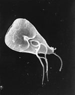 Giardia intestinalis (eukaryote) Giardia intestinalis (eukaryote) | ||||||
| Method | X-RAY DIFFRACTION / SYNCHROTRON / MAD / Resolution: 1.9 Å | ||||||
 Authors Authors | Weeratunga, S. / Hofmann, A. | ||||||
 Citation Citation | Journal: J.Mol.Biol. / Year: 2012 Title: Alpha-1 giardin is an annexin with highly unusual calcium-regulated mechanisms Authors: Weeratunga, S.K. / Osman, A. / Hu, N.-J. / Wang, C.K. / Mason, L. / Svard, S. / Hope, G. / Jones, M.K. / Hofmann, A. | ||||||
| History |
|
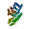
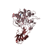
- Structure visualization
| Structure viewer | Molecule: MolmilJmol/JSmol |
|---|
- Downloads & links
-Download
| PDBx/mmCIF format | 4evf.cif.gz | 76.3 KB | Display | PDBx/mmCIF format |
|---|---|---|---|---|
| PDB format | pdb4evf.ent.gz | 56.8 KB | Display | PDB format |
| PDBx/mmJSON format | 4evf.json.gz | Tree view | PDBx/mmJSON format | |
| Others |  Other downloads Other downloads |
-Validation report
| Arichive directory | https://data.pdbj.org/pub/pdb/validation_reports/ev/4evfftp://data.pdbj.org/pub/pdb/validation_reports/ev/4evf | HTTPS FTP |
|---|
-Related structure data


-Links
 PDBj
PDBj- Assembly
| Deposited unit | 
| ||||||||
|---|---|---|---|---|---|---|---|---|---|
| 1 |
| ||||||||
| Unit cell |
|
-Components
| #1: Protein | Mass: 33941.914 Da / Num. of mol.: 1 Source method: isolated from a genetically manipulated source Source: (gene. exp.) Giardia intestinalis (eukaryote) / Plasmid: pRSET_6c / Production host:  Escherichia coli (E. coli) / Strain (production host): BL21(DE3) / References: UniProt: P17063 Escherichia coli (E. coli) / Strain (production host): BL21(DE3) / References: UniProt: P17063 |
|---|---|
| #2: Water | ChemComp-HOH / Water Mass: 18.015 Da / Num. of mol.: 286 / Source method: isolated from a natural source / Formula: H2O Mass: 18.015 Da / Num. of mol.: 286 / Source method: isolated from a natural source / Formula: H2O |
-Experimental details
-Experiment
| Experiment | Method: X-RAY DIFFRACTION / Number of used crystals: 2 |
|---|
- Sample preparation
| Crystal |
| ||||||||||||
|---|---|---|---|---|---|---|---|---|---|---|---|---|---|
| Crystal grow | Temperature: 289 K / Method: vapor diffusion, hanging drop / pH: 8 Details: 0.1M MgCl2, 30% PEG 2000, 20mM KH2PO4, pH 8.0, VAPOR DIFFUSION, HANGING DROP, temperature 289K |
-Data collection
| Diffraction |
| |||||||||||||||
|---|---|---|---|---|---|---|---|---|---|---|---|---|---|---|---|---|
| Diffraction source |
| |||||||||||||||
| Detector |
| |||||||||||||||
| Radiation |
| |||||||||||||||
| Radiation wavelength |
| |||||||||||||||
| Reflection | Resolution: 1.9→50 Å / Num. obs: 47848 / % possible obs: 0.983 % / Redundancy: 5.1 % / Biso Wilson estimate: 25.7 Å2 / Rsym value: 0.123 | |||||||||||||||
| Reflection shell | Resolution: 1.9→2 Å / Redundancy: 4.8 % / Num. unique all: 3386 / Rsym value: 0.382 / % possible all: 97.2 |


- Processing
| Software |
| |||||||||||||||||||||||||||||||||||||||||||||||||||||||||||||||||||||||||||||||||||||||||||||||||||||||||||||||||||||||
|---|---|---|---|---|---|---|---|---|---|---|---|---|---|---|---|---|---|---|---|---|---|---|---|---|---|---|---|---|---|---|---|---|---|---|---|---|---|---|---|---|---|---|---|---|---|---|---|---|---|---|---|---|---|---|---|---|---|---|---|---|---|---|---|---|---|---|---|---|---|---|---|---|---|---|---|---|---|---|---|---|---|---|---|---|---|---|---|---|---|---|---|---|---|---|---|---|---|---|---|---|---|---|---|---|---|---|---|---|---|---|---|---|---|---|---|---|---|---|---|---|
| Refinement | Method to determine structure: MAD / Resolution: 1.9→24.826 Å / Occupancy max: 1 / Occupancy min: 0 / SU ML: 0.29 / σ(F): 1.39 / Phase error: 25.92 / Stereochemistry target values: ML / Details: THE FRIEDEL PAIRS WERE USED FOR PHASING.
| |||||||||||||||||||||||||||||||||||||||||||||||||||||||||||||||||||||||||||||||||||||||||||||||||||||||||||||||||||||||
| Solvent computation | Shrinkage radii: 0.72 Å / VDW probe radii: 1 Å / Solvent model: FLAT BULK SOLVENT MODEL / Bsol: 46.106 Å2 / ksol: 0.377 e/Å3 | |||||||||||||||||||||||||||||||||||||||||||||||||||||||||||||||||||||||||||||||||||||||||||||||||||||||||||||||||||||||
| Displacement parameters | Biso max: 55.04 Å2 / Biso mean: 26.3783 Å2 / Biso min: 13.13 Å2
| |||||||||||||||||||||||||||||||||||||||||||||||||||||||||||||||||||||||||||||||||||||||||||||||||||||||||||||||||||||||
| Refinement step | Cycle: LAST / Resolution: 1.9→24.826 Å
| |||||||||||||||||||||||||||||||||||||||||||||||||||||||||||||||||||||||||||||||||||||||||||||||||||||||||||||||||||||||
| Refine LS restraints |
| |||||||||||||||||||||||||||||||||||||||||||||||||||||||||||||||||||||||||||||||||||||||||||||||||||||||||||||||||||||||
| LS refinement shell | Refine-ID: X-RAY DIFFRACTION / Total num. of bins used: 16
|
