 Movie
Movie Controller

[English] 日本語
 Yorodumi
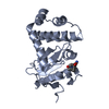
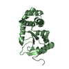
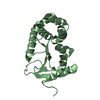
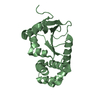
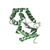
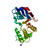
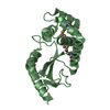
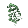
- PDB-4eto: Structure of S100A4 in complex with non-muscle myosin-IIA peptide -
+ Open data

- Basic information
| Entry | Database: PDB / ID: 4eto | ||||||
|---|---|---|---|---|---|---|---|
| Title | Structure of S100A4 in complex with non-muscle myosin-IIA peptide | ||||||
 Components Components |
| ||||||
 Keywords Keywords | METAL BINDING PROTEIN /  CALCIUM-BINDING PROTEIN / EF-hand / Structural Genomics / PSI-Biology / Protein Structure Initiative / New York Structural Genomics Research Consortium / NYSGRC / Atoms-to-Animals: The Immune Function Network / IFN CALCIUM-BINDING PROTEIN / EF-hand / Structural Genomics / PSI-Biology / Protein Structure Initiative / New York Structural Genomics Research Consortium / NYSGRC / Atoms-to-Animals: The Immune Function Network / IFN | ||||||
| Function / homology |  Function and homology information Function and homology informationnegative regulation of actin filament severing / uropod organization / regulation of plasma membrane repair / cortical granule exocytosis / cytokinetic process / establishment of meiotic spindle localization / myosin II filament / establishment of T cell polarity / cortical granule / actomyosin contractile ring ...negative regulation of actin filament severing / uropod organization / regulation of plasma membrane repair / cortical granule exocytosis / cytokinetic process / establishment of meiotic spindle localization / myosin II filament / establishment of T cell polarity / cortical granule / actomyosin contractile ring / positive regulation of protein processing in phagocytic vesicle / regulated exocytosis / uropod / actin filament-based movement / blood vessel endothelial cell migration / meiotic spindle organization / RAGE receptor binding / actomyosin / plasma membrane repair / lysosome localization / myosin filament / myoblast fusion / actomyosin structure organization / myosin II complex / RHO GTPases Activate ROCKs / RHO GTPases activate CIT / Sema4D induced cell migration and growth-cone collapse / Sensory processing of sound by outer hair cells of the cochlea / Sensory processing of sound by inner hair cells of the cochlea / CD163 mediating an anti-inflammatory response / microfilament motor activity / platelet formation / phagocytosis, engulfment / leukocyte migration / EPHA-mediated growth cone collapse / endodermal cell differentiation / chemoattractant activity / cell leading edge / RHO GTPases activate PAKs / cleavage furrow / cytoskeletal motor activity / monocyte differentiation / brush border / membrane protein ectodomain proteolysis / transition metal ion binding / Signaling by ALK fusions and activated point mutants / immunological synapse / epithelial to mesenchymal transition / stress fiber / protein-membrane adaptor activity / RHO GTPases activate PKNs / ruffle / integrin-mediated signaling pathway / Translocation of SLC2A4 (GLUT4) to the plasma membrane / ADP binding / FCGR3A-mediated phagocytosis / adherens junction / neuromuscular junction / cytoplasmic side of plasma membrane / spindle / Regulation of actin dynamics for phagocytic cup formation / platelet aggregation / calcium-dependent protein binding / actin filament binding / actin cytoskeleton / integrin binding / protein transport / actin binding / regulation of cell shape / actin cytoskeleton organization / angiogenesis / collagen-containing extracellular matrix / positive regulation of canonical NF-kappaB signal transduction / in utero embryonic development / nuclear body / calmodulin binding / cadherin binding / protein domain specific binding / focal adhesion / calcium ion binding / perinuclear region of cytoplasm / protein homodimerization activity / protein-containing complex / extracellular space / RNA binding / extracellular exosome / extracellular region / nucleoplasm / ATP binding / membrane / identical protein binding / nucleus / plasma membrane / cytosol / cytoplasmSimilarity search - Function | ||||||
| Biological species |  Homo sapiens (human) Homo sapiens (human) | ||||||
| Method | X-RAY DIFFRACTION / SYNCHROTRON / MOLECULAR REPLACEMENT / Resolution: 1.54 Å | ||||||
 Authors Authors | Ramagopal, U.A. / Dulyaninova, N.G. / Kumar, P.R. / Almo, S.C. / Bresnick, A.R. / New York Structural Genomics Research Consortium (NYSGRC) / Atoms-to-Animals: The Immune Function Network (IFN) | ||||||
 Citation Citation | Journal: To be Published Title: Structure of S100A4 with bound peptide P Authors: Ramagopal, U.A. / Dulyaninova, N.G. / Kumar, P.R. / Almo, S.C. / Bresnick, A.R. | ||||||
| History |
|
- Structure visualization
| Structure viewer | Molecule: MolmilJmol/JSmol |
|---|
- Downloads & links
-Download
| PDBx/mmCIF format | 4eto.cif.gz | 56.5 KB | Display | PDBx/mmCIF format |
|---|---|---|---|---|
| PDB format | pdb4eto.ent.gz | 39.7 KB | Display | PDB format |
| PDBx/mmJSON format | 4eto.json.gz | Tree view | PDBx/mmJSON format | |
| Others |  Other downloads Other downloads |
-Validation report
| Arichive directory | https://data.pdbj.org/pub/pdb/validation_reports/et/4etoftp://data.pdbj.org/pub/pdb/validation_reports/et/4eto | HTTPS FTP |
|---|
-Related structure data
| Related structure data |  2q91S S: Starting model for refinement |
|---|---|
| Similar structure data | |
| Other databases |


-Links
 PDBj
PDBj


- Assembly
| Deposited unit | 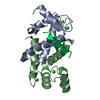
| ||||||||
|---|---|---|---|---|---|---|---|---|---|
| 1 |
| ||||||||
| Unit cell |
| ||||||||
| Details | dimer bound to a peptide |
-Components
| #1: Protein | / Calvasculin / Metastasin / Placental calcium-binding protein / Protein Mts1 / S100 calcium-binding protein A4 Mass: 10763.358 Da / Num. of mol.: 2 / Fragment: UNP residues 1-93 Source method: isolated from a genetically manipulated source Source: (gene. exp.) Homo sapiens (human) / Gene: CAPL, MTS1, S100A4 / Plasmid: PET23A) / Production host:  Escherichia coli (E. coli) / Strain (production host): BL(DE3) / References: UniProt: P26447 Escherichia coli (E. coli) / Strain (production host): BL(DE3) / References: UniProt: P26447#2: Protein/peptide | | / Cellular myosin heavy chain / type A / Myosin heavy chain 9 / Myosin heavy chain / non-muscle IIa / ...Cellular myosin heavy chain / type A / Myosin heavy chain 9 / Myosin heavy chain / non-muscle IIa / Non-muscle myosin heavy chain A / NMMHC-A / Non-muscle myosin heavy chain IIa / NMMHC II-a / NMMHC-IIAMass: 1922.239 Da / Num. of mol.: 1 / Fragment: Coiled coil region residues 1908-1923 Source method: isolated from a genetically manipulated source Source: (gene. exp.) Homo sapiens (human) / Gene: MYH9 / References: UniProt: P35579#3: Chemical | ChemComp-CA /   Mass: 40.078 Da / Num. of mol.: 4 / Source method: obtained synthetically / Formula: Ca Mass: 40.078 Da / Num. of mol.: 4 / Source method: obtained synthetically / Formula: Ca#4: Water | ChemComp-HOH / | Water Mass: 18.015 Da / Num. of mol.: 86 / Source method: isolated from a natural source / Formula: H2O Mass: 18.015 Da / Num. of mol.: 86 / Source method: isolated from a natural source / Formula: H2O |
|---|
-Experimental details
-Experiment
| Experiment | Method: X-RAY DIFFRACTION / Number of used crystals: 1 |
|---|
- Sample preparation
| Crystal | Density Matthews: 1.8 Å3/Da / Density % sol: 31.74 % |
|---|---|
| Crystal grow | Temperature: 298 K / Method: vapor diffusion, sitting drop / pH: 7.5 Details: 0.1 M HEPES pH 7.0, 30% (v/v) Jeffamine ED-2001, Vapor diffusion, Sitting drop, temperature 298K |
-Data collection
| Diffraction | Mean temperature: 100 K | |||||||||||||||||||||||||||||||||||||||||||||||||||||||||||||||||||||||||||||||||||||||||||||||||||||||||||||||||||||||||||||||||||||||||||||||||||
|---|---|---|---|---|---|---|---|---|---|---|---|---|---|---|---|---|---|---|---|---|---|---|---|---|---|---|---|---|---|---|---|---|---|---|---|---|---|---|---|---|---|---|---|---|---|---|---|---|---|---|---|---|---|---|---|---|---|---|---|---|---|---|---|---|---|---|---|---|---|---|---|---|---|---|---|---|---|---|---|---|---|---|---|---|---|---|---|---|---|---|---|---|---|---|---|---|---|---|---|---|---|---|---|---|---|---|---|---|---|---|---|---|---|---|---|---|---|---|---|---|---|---|---|---|---|---|---|---|---|---|---|---|---|---|---|---|---|---|---|---|---|---|---|---|---|---|---|---|
| Diffraction source | Source: SYNCHROTRON / Site: NSLS  / Beamline: X4A / Wavelength: 0.9793 Å / Beamline: X4A / Wavelength: 0.9793 Å | |||||||||||||||||||||||||||||||||||||||||||||||||||||||||||||||||||||||||||||||||||||||||||||||||||||||||||||||||||||||||||||||||||||||||||||||||||
| Detector | Type: ADSC QUANTUM 210 / Detector: CCD / Date: Feb 17, 2011 | |||||||||||||||||||||||||||||||||||||||||||||||||||||||||||||||||||||||||||||||||||||||||||||||||||||||||||||||||||||||||||||||||||||||||||||||||||
| Radiation | Protocol: SINGLE WAVELENGTH / Scattering type: x-ray | |||||||||||||||||||||||||||||||||||||||||||||||||||||||||||||||||||||||||||||||||||||||||||||||||||||||||||||||||||||||||||||||||||||||||||||||||||
| Radiation wavelength | Wavelength: 0.9793 Å / Relative weight: 1 | |||||||||||||||||||||||||||||||||||||||||||||||||||||||||||||||||||||||||||||||||||||||||||||||||||||||||||||||||||||||||||||||||||||||||||||||||||
| Reflection | Resolution: 1.54→50 Å / Num. obs: 24461 / % possible obs: 99.6 % / Redundancy: 3.6 % / Rmerge(I) obs: 0.063 / Χ2: 1.734 / Net I/σ(I): 9.9 | |||||||||||||||||||||||||||||||||||||||||||||||||||||||||||||||||||||||||||||||||||||||||||||||||||||||||||||||||||||||||||||||||||||||||||||||||||
| Reflection shell |
|
- Processing
| Software |
| |||||||||||||||||||||||||||||||||||||||||||||||||||||||||||||||||
|---|---|---|---|---|---|---|---|---|---|---|---|---|---|---|---|---|---|---|---|---|---|---|---|---|---|---|---|---|---|---|---|---|---|---|---|---|---|---|---|---|---|---|---|---|---|---|---|---|---|---|---|---|---|---|---|---|---|---|---|---|---|---|---|---|---|---|
| Refinement | Method to determine structure: MOLECULAR REPLACEMENT Starting model: 2Q91 Resolution: 1.54→45.99 Å / Cor.coef. Fo:Fc: 0.95 / Cor.coef. Fo:Fc free: 0.927 / WRfactor Rfree: 0.2494 / WRfactor Rwork: 0.2093 / Occupancy max: 1 / Occupancy min: 0.3 / FOM work R set: 0.8274 / SU B: 1.913 / SU ML: 0.07 / SU R Cruickshank DPI: 0.1053 / SU Rfree: 0.1066 / Cross valid method: THROUGHOUT / σ(F): 0 / ESU R: 0.105 / ESU R Free: 0.107 / Stereochemistry target values: MAXIMUM LIKELIHOOD Details: HYDROGENS HAVE BEEN ADDED IN THE RIDING POSITIONS U VALUES : REFINED INDIVIDUALLY
| |||||||||||||||||||||||||||||||||||||||||||||||||||||||||||||||||
| Solvent computation | Ion probe radii: 0.8 Å / Shrinkage radii: 0.8 Å / VDW probe radii: 1.4 Å / Solvent model: MASK | |||||||||||||||||||||||||||||||||||||||||||||||||||||||||||||||||
| Displacement parameters | Biso max: 50.68 Å2 / Biso mean: 20.1365 Å2 / Biso min: 6.21 Å2
| |||||||||||||||||||||||||||||||||||||||||||||||||||||||||||||||||
| Refinement step | Cycle: LAST / Resolution: 1.54→45.99 Å
| |||||||||||||||||||||||||||||||||||||||||||||||||||||||||||||||||
| Refine LS restraints |
| |||||||||||||||||||||||||||||||||||||||||||||||||||||||||||||||||
| LS refinement shell | Resolution: 1.54→1.58 Å / Total num. of bins used: 20
|
