 Movie
Movie Controller


+ Open data

- Basic information
| Entry | Database: PDB / ID: 4er0 | ||||||
|---|---|---|---|---|---|---|---|
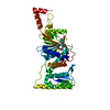
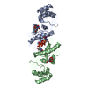
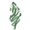
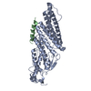
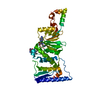
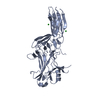
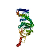
| Title | Crystal Structure of human DOT1L in complex with inhibitor FED1 | ||||||
 Components Components | Histone-lysine N-methyltransferase, H3 lysine-79 specific Histone methyltransferase Histone methyltransferase | ||||||
 Keywords Keywords | Transferase/Transferase Inhibitor / histone / methyltransferase / epigenetics / Transferase-Transferase Inhibitor complex / Structural Genomics / Structural Genomics Consortium / SGC | ||||||
| Function / homology |  Function and homology information Function and homology informationhistone H3K79 trimethyltransferase activity / [histone H3]-lysine79 N-trimethyltransferase / histone H3K79 methyltransferase activity / regulation of transcription regulatory region DNA binding / histone H3 methyltransferase activity / regulation of receptor signaling pathway via JAK-STAT / histone methyltransferase activity / heterochromatin formation / telomere organization / DNA damage checkpoint signaling ...histone H3K79 trimethyltransferase activity / [histone H3]-lysine79 N-trimethyltransferase / histone H3K79 methyltransferase activity / regulation of transcription regulatory region DNA binding / histone H3 methyltransferase activity / regulation of receptor signaling pathway via JAK-STAT / histone methyltransferase activity / heterochromatin formation / telomere organization / DNA damage checkpoint signaling / PKMTs methylate histone lysines / gene expression / methylation / RNA polymerase II-specific DNA-binding transcription factor binding / nucleic acid binding / transcription coactivator activity / intracellular membrane-bounded organelle / DNA repair / positive regulation of cell population proliferation / positive regulation of transcription by RNA polymerase II / protein-containing complex / DNA binding / nucleoplasm / nucleus / cytoplasmSimilarity search - Function | ||||||
| Biological species |  Homo sapiens (human) Homo sapiens (human) | ||||||
| Method | X-RAY DIFFRACTION / SYNCHROTRON / FOURIER SYNTHESIS / Resolution: 2.5 Å | ||||||
 Authors Authors | Wernimont, A.K. / Tempel, W. / Yu, W. / Li, Y. / Nguyen, K.T. / Federation, A. / Marineau, J. / Qi, J. / Vedadi, M. / Bradner, J.E. ...Wernimont, A.K. / Tempel, W. / Yu, W. / Li, Y. / Nguyen, K.T. / Federation, A. / Marineau, J. / Qi, J. / Vedadi, M. / Bradner, J.E. / Schapira, M. / Arrowsmith, C.H. / Edwards, A.M. / Bountra, C. / Brown, P.J. / Structural Genomics Consortium (SGC) | ||||||
 Citation Citation | Journal: Nat Commun / Year: 2012 Title: Catalytic site remodelling of the DOT1L methyltransferase by selective inhibitors. Authors: Yu, W. / Chory, E.J. / Wernimont, A.K. / Tempel, W. / Scopton, A. / Federation, A. / Marineau, J.J. / Qi, J. / Barsyte-Lovejoy, D. / Yi, J. / Marcellus, R. / Iacob, R.E. / Engen, J.R. / ...Authors: Yu, W. / Chory, E.J. / Wernimont, A.K. / Tempel, W. / Scopton, A. / Federation, A. / Marineau, J.J. / Qi, J. / Barsyte-Lovejoy, D. / Yi, J. / Marcellus, R. / Iacob, R.E. / Engen, J.R. / Griffin, C. / Aman, A. / Wienholds, E. / Li, F. / Pineda, J. / Estiu, G. / Shatseva, T. / Hajian, T. / Al-Awar, R. / Dick, J.E. / Vedadi, M. / Brown, P.J. / Arrowsmith, C.H. / Bradner, J.E. / Schapira, M. | ||||||
| History |
|
- Structure visualization
| Structure viewer | Molecule: MolmilJmol/JSmol |
|---|
- Downloads & links
-Download
| PDBx/mmCIF format | 4er0.cif.gz | 79.2 KB | Display | PDBx/mmCIF format |
|---|---|---|---|---|
| PDB format | pdb4er0.ent.gz | 57.2 KB | Display | PDB format |
| PDBx/mmJSON format | 4er0.json.gz | Tree view | PDBx/mmJSON format | |
| Others |  Other downloads Other downloads |
-Validation report
| Arichive directory | https://data.pdbj.org/pub/pdb/validation_reports/er/4er0ftp://data.pdbj.org/pub/pdb/validation_reports/er/4er0 | HTTPS FTP |
|---|
-Related structure data
| Related structure data |  3uwpC  4eqzC  4er3C  4er5C  4er6C  4er7C C: citing same article ( |
|---|---|
| Similar structure data |


-Links
 PDBj
PDBj
- Assembly
| Deposited unit | 
| ||||||||
|---|---|---|---|---|---|---|---|---|---|
| 1 |
| ||||||||
| Unit cell |
| ||||||||
| Details | AUTHORS STATE THAT THE BIOLOGICAL MOLECULE IS UNKNOWN. |
-Components
| #1: Protein | Histone methyltransferase / DOT1-like protein / Histone H3-K79 methyltransferase / H3-K79-HMTase / Lysine N-methyltransferase 4 Mass: 47980.688 Da / Num. of mol.: 1 Source method: isolated from a genetically manipulated source Source: (gene. exp.) Homo sapiens (human) / Gene: DOT1L, KIAA1814, KMT4 / Plasmid: pET28-MHL / Production host:  Escherichia coli (E. coli) / Strain (production host): BL21 V2r Prare2 Escherichia coli (E. coli) / Strain (production host): BL21 V2r Prare2References: UniProt: Q8TEK3, histone-lysine N-methyltransferase | ||
|---|---|---|---|
| #2: Chemical | ChemComp-AW1 /   Mass: 540.658 Da / Num. of mol.: 1 / Source method: obtained synthetically / Formula: C27H40N8O4 Mass: 540.658 Da / Num. of mol.: 1 / Source method: obtained synthetically / Formula: C27H40N8O4 | ||
| #3: Chemical |   Num. of mol.: 3 / Source method: obtained synthetically Num. of mol.: 3 / Source method: obtained synthetically#4: Water | ChemComp-HOH / | Water Mass: 18.015 Da / Num. of mol.: 6 / Source method: isolated from a natural source / Formula: H2O Mass: 18.015 Da / Num. of mol.: 6 / Source method: isolated from a natural source / Formula: H2O |
-Experimental details
-Experiment
| Experiment | Method: X-RAY DIFFRACTION / Number of used crystals: 1 |
|---|
- Sample preparation
| Crystal | Density Matthews: 3.61 Å3/Da / Density % sol: 65.9 % |
|---|---|
| Crystal grow | Temperature: 293 K / Method: vapor diffusion, hanging drop / pH: 4.5 Details: 3.5 M NaFormate, 0.1 M NaAcet, 0.5 mM inhibitor in H2O, pH 4.5, VAPOR DIFFUSION, HANGING DROP, temperature 293K |
-Data collection
| Diffraction | Mean temperature: 100 K | ||||||||||||||||||||||||||||||||||||||||||||||||||||||||||||||||||||||||||||||||||||||||||||||||||||||||||||||||||||||||||||||
|---|---|---|---|---|---|---|---|---|---|---|---|---|---|---|---|---|---|---|---|---|---|---|---|---|---|---|---|---|---|---|---|---|---|---|---|---|---|---|---|---|---|---|---|---|---|---|---|---|---|---|---|---|---|---|---|---|---|---|---|---|---|---|---|---|---|---|---|---|---|---|---|---|---|---|---|---|---|---|---|---|---|---|---|---|---|---|---|---|---|---|---|---|---|---|---|---|---|---|---|---|---|---|---|---|---|---|---|---|---|---|---|---|---|---|---|---|---|---|---|---|---|---|---|---|---|---|---|
| Diffraction source | Source: SYNCHROTRON / Site: APS  / Beamline: 19-ID / Wavelength: 0.97907 Å / Beamline: 19-ID / Wavelength: 0.97907 Å | ||||||||||||||||||||||||||||||||||||||||||||||||||||||||||||||||||||||||||||||||||||||||||||||||||||||||||||||||||||||||||||||
| Detector | Type: ADSC QUANTUM 315 / Detector: CCD / Date: Nov 30, 2011 | ||||||||||||||||||||||||||||||||||||||||||||||||||||||||||||||||||||||||||||||||||||||||||||||||||||||||||||||||||||||||||||||
| Radiation | Protocol: SINGLE WAVELENGTH / Monochromatic (M) / Laue (L): M / Scattering type: x-ray | ||||||||||||||||||||||||||||||||||||||||||||||||||||||||||||||||||||||||||||||||||||||||||||||||||||||||||||||||||||||||||||||
| Radiation wavelength | Wavelength: 0.97907 Å / Relative weight: 1 | ||||||||||||||||||||||||||||||||||||||||||||||||||||||||||||||||||||||||||||||||||||||||||||||||||||||||||||||||||||||||||||||
| Reflection | Resolution: 2.5→40 Å / Num. all: 24065 / Num. obs: 24065 / % possible obs: 100 % / Observed criterion σ(F): 0 / Observed criterion σ(I): 0 / Redundancy: 11.1 % / Biso Wilson estimate: 63 Å2 / Rmerge(I) obs: 0.07 / Χ2: 0.74 / Net I/σ(I): 7.2 | ||||||||||||||||||||||||||||||||||||||||||||||||||||||||||||||||||||||||||||||||||||||||||||||||||||||||||||||||||||||||||||||
| Reflection shell | Diffraction-ID: 1 / % possible all: 100
|
- Processing
| Software |
| |||||||||||||||||||||||||||||||||||||||||||||||||||||||||||||||||||||||||||||||||||||
|---|---|---|---|---|---|---|---|---|---|---|---|---|---|---|---|---|---|---|---|---|---|---|---|---|---|---|---|---|---|---|---|---|---|---|---|---|---|---|---|---|---|---|---|---|---|---|---|---|---|---|---|---|---|---|---|---|---|---|---|---|---|---|---|---|---|---|---|---|---|---|---|---|---|---|---|---|---|---|---|---|---|---|---|---|---|---|
| Refinement | Method to determine structure: FOURIER SYNTHESIS / Resolution: 2.5→35 Å / Cor.coef. Fo:Fc: 0.932 / Cor.coef. Fo:Fc free: 0.908 / WRfactor Rfree: 0.2672 / WRfactor Rwork: 0.2286 / Occupancy max: 1 / Occupancy min: 0.3 / FOM work R set: 0.8027 / SU B: 7.553 / SU ML: 0.172 / SU R Cruickshank DPI: 0.2661 / SU Rfree: 0.2289 / Cross valid method: THROUGHOUT / σ(F): 0 / ESU R: 0.266 / ESU R Free: 0.229 / Stereochemistry target values: MAXIMUM LIKELIHOOD / Details: HYDROGENS HAVE BEEN ADDED IN THE RIDING POSITIONS
| |||||||||||||||||||||||||||||||||||||||||||||||||||||||||||||||||||||||||||||||||||||
| Solvent computation | Ion probe radii: 0.8 Å / Shrinkage radii: 0.8 Å / VDW probe radii: 1.4 Å / Solvent model: MASK | |||||||||||||||||||||||||||||||||||||||||||||||||||||||||||||||||||||||||||||||||||||
| Displacement parameters | Biso max: 89.83 Å2 / Biso mean: 52.6636 Å2 / Biso min: 26.85 Å2
| |||||||||||||||||||||||||||||||||||||||||||||||||||||||||||||||||||||||||||||||||||||
| Refinement step | Cycle: LAST / Resolution: 2.5→35 Å
| |||||||||||||||||||||||||||||||||||||||||||||||||||||||||||||||||||||||||||||||||||||
| Refine LS restraints |
| |||||||||||||||||||||||||||||||||||||||||||||||||||||||||||||||||||||||||||||||||||||
| LS refinement shell | Resolution: 2.5→2.565 Å / Total num. of bins used: 20
|
