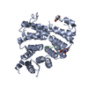
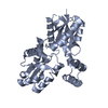
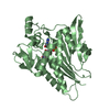
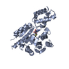
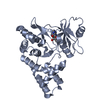
PROTEIN BINDING/TRANSPORT PROTEIN / Accessory protein / Tetratricopeptide repeat / TPR / Accessory Protein for HCN channels / HCN / PROTEIN BINDING-TRANSPORT PROTEIN complex
Function / homology
Function and homology information
intracellularly cyclic nucleotide-activated monoatomic cation channel activity / peroxisome matrix targeting signal-1 binding / HCN channels / peroxisome targeting sequence binding / small GTPase binding => GO:0031267 / positive regulation of corticotropin secretion / protein import into peroxisome matrix, docking / HCN channel complex / intracellular cyclic nucleotide activated cation channel complex / regulated exocytosis ...intracellularly cyclic nucleotide-activated monoatomic cation channel activity / peroxisome matrix targeting signal-1 binding / HCN channels / peroxisome targeting sequence binding / small GTPase binding => GO:0031267 / positive regulation of corticotropin secretion / protein import into peroxisome matrix, docking / HCN channel complex / intracellular cyclic nucleotide activated cation channel complex / regulated exocytosis / cell tip / intracellularly cAMP-activated cation channel activity / cellular response to cGMP / maintenance of protein location / sodium ion import across plasma membrane / peroxisomal membrane / voltage-gated sodium channel activity / regulation of membrane depolarization / potassium ion import across plasma membrane / voltage-gated potassium channel activity / sodium ion transmembrane transport / regulation of cAMP-mediated signaling / cAMP binding / cellular response to cAMP / potassium ion transmembrane transport / somatodendritic compartment / dendrite membrane / dendritic shaft / regulation of membrane potential / PDZ domain binding / small GTPase binding / molecular adaptor activity / receptor complex / axon / neuronal cell body / dendrite / protein-containing complex binding / perinuclear region of cytoplasm / membrane / identical protein binding / plasma membrane / cytosol / cytoplasm Similarity search - Function
PEX5-related, vertebrates / PEX5/PEX5L / Ion transport N-terminal / Ion transport protein N-terminal / : / Potassium channel, voltage-dependent, EAG/ELK/ERG / Tetratricopeptide repeat / Cyclic nucleotide-binding domain signature 1. / Cyclic nucleotide-binding, conserved site / Tetratricopeptide repeat domain ...PEX5-related, vertebrates / PEX5/PEX5L / Ion transport N-terminal / Ion transport protein N-terminal / : / Potassium channel, voltage-dependent, EAG/ELK/ERG / Tetratricopeptide repeat / Cyclic nucleotide-binding domain signature 1. / Cyclic nucleotide-binding, conserved site / Tetratricopeptide repeat domain / Tetratricopeptide repeat / Cyclic nucleotide-monophosphate binding domain / Cyclic nucleotide-binding domain / cAMP/cGMP binding motif profile. / Cyclic nucleotide-binding domain / Cyclic nucleotide-binding domain superfamily / TPR repeat region circular profile. / TPR repeat profile. / Tetratricopeptide repeats / Tetratricopeptide repeat / RmlC-like jelly roll fold / Serine Threonine Protein Phosphatase 5, Tetratricopeptide repeat / Ion transport domain / Ion transport protein / Alpha Horseshoe / Tetratricopeptide-like helical domain superfamily / Mainly Alpha Similarity search - Domain/homology
In the structure databanks used in Yorodumi, some data are registered as the other names, "COVID-19 virus" and "2019-nCoV". Here are the details of the virus and the list of structure data.
Jan 31, 2019. EMDB accession codes are about to change! (news from PDBe EMDB page)
EMDB accession codes are about to change! (news from PDBe EMDB page)
The allocation of 4 digits for EMDB accession codes will soon come to an end. Whilst these codes will remain in use, new EMDB accession codes will include an additional digit and will expand incrementally as the available range of codes is exhausted. The current 4-digit format prefixed with “EMD-” (i.e. EMD-XXXX) will advance to a 5-digit format (i.e. EMD-XXXXX), and so on. It is currently estimated that the 4-digit codes will be depleted around Spring 2019, at which point the 5-digit format will come into force.
The EM Navigator/Yorodumi systems omit the EMD- prefix.
Related info.:Q: What is EMD? / ID/Accession-code notation in Yorodumi/EM Navigator
Yorodumi is a browser for structure data from EMDB, PDB, SASBDB, etc.
This page is also the successor to EM Navigator detail page, and also detail information page/front-end page for Omokage search.
The word "yorodu" (or yorozu) is an old Japanese word meaning "ten thousand". "mi" (miru) is to see.
Related info.:EMDB / PDB / SASBDB / Comparison of 3 databanks / Yorodumi Search / Aug 31, 2016. New EM Navigator & Yorodumi / Yorodumi Papers / Jmol/JSmol / Function and homology information / Changes in new EM Navigator and Yorodumi
 Movie
Movie Controller

 Yorodumi
 Open data
 Basic information
 Components
 Keywords
 Tetratricopeptide repeat / TPR / Accessory Protein for HCN channels / HCN / PROTEIN BINDING-TRANSPORT PROTEIN complex
 Function and homology information
 Authors
 Citation
 Structure visualization
 Downloads & links
 Other downloads


 PDBj
PDBj


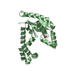
 Assembly
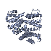

 Sample preparation
 / Beamline: 8.2.1 / Wavelength: 0.9999 Å
 Processing