 Movie
Movie Controller

[English] 日本語
 Yorodumi
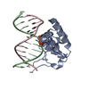
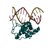
- PDB-4enk: Crystal structure of S. pombe Atl1 in complex with damaged DNA co... -
+ Open data

- Basic information
| Entry | Database: PDB / ID: 4enk | ||||||
|---|---|---|---|---|---|---|---|
| Title | Crystal structure of S. pombe Atl1 in complex with damaged DNA containing O6-propylguanine | ||||||
 Components Components |
| ||||||
 Keywords Keywords | DNA BINDING PROTEIN/DNA / Alkyltransferase /  DNA repair / nucleotide excision repair / NER / base repair / DNA / DNA Damage / Guanine / Alkylation / DNA BINDING PROTEIN-DNA complex DNA repair / nucleotide excision repair / NER / base repair / DNA / DNA Damage / Guanine / Alkylation / DNA BINDING PROTEIN-DNA complex | ||||||
| Function / homology |  Function and homology information Function and homology informationO6-alkylguanine-DNA binding / global genome nucleotide-excision repair / transcription-coupled nucleotide-excision repair / catalytic activity / damaged DNA binding / nucleus / cytosolSimilarity search - Function | ||||||
| Biological species |  Schizosaccharomyces pombe (fission yeast) Schizosaccharomyces pombe (fission yeast)synthetic construct (others) | ||||||
| Method | X-RAY DIFFRACTION / SYNCHROTRON / MOLECULAR REPLACEMENT / Resolution: 3.0445 Å | ||||||
 Authors Authors | Tubbs, J.L. / Arvai, A.S. / Tainer, J.A. | ||||||
 Citation Citation | Journal: Mol.Cell / Year: 2012 Title: Atl1 Regulates Choice between Global Genome and Transcription-Coupled Repair of O(6)-Alkylguanines. Authors: Latypov, V.F. / Tubbs, J.L. / Watson, A.J. / Marriott, A.S. / McGown, G. / Thorncroft, M. / Wilkinson, O.J. / Senthong, P. / Butt, A. / Arvai, A.S. / Millington, C.L. / Povey, A.C. / ...Authors: Latypov, V.F. / Tubbs, J.L. / Watson, A.J. / Marriott, A.S. / McGown, G. / Thorncroft, M. / Wilkinson, O.J. / Senthong, P. / Butt, A. / Arvai, A.S. / Millington, C.L. / Povey, A.C. / Williams, D.M. / Santibanez-Koref, M.F. / Tainer, J.A. / Margison, G.P. | ||||||
| History |
|
- Structure visualization
| Structure viewer | Molecule: MolmilJmol/JSmol |
|---|
- Downloads & links
-Download
| PDBx/mmCIF format | 4enk.cif.gz | 53.8 KB | Display | PDBx/mmCIF format |
|---|---|---|---|---|
| PDB format | pdb4enk.ent.gz | 35.2 KB | Display | PDB format |
| PDBx/mmJSON format | 4enk.json.gz | Tree view | PDBx/mmJSON format | |
| Others |  Other downloads Other downloads |
-Validation report
| Arichive directory | https://data.pdbj.org/pub/pdb/validation_reports/en/4enkftp://data.pdbj.org/pub/pdb/validation_reports/en/4enk | HTTPS FTP |
|---|
-Related structure data
| Related structure data |  4enjC  4enmC 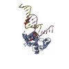 4ennC 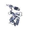 3gvaS C: citing same article ( S: Starting model for refinement |
|---|---|
| Similar structure data |


-Links
 PDBj
PDBj


- Assembly
| Deposited unit | 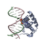
| ||||||||
|---|---|---|---|---|---|---|---|---|---|
| 1 |
| ||||||||
| Unit cell |
|
-Components
| #1: Protein | Mass: 13662.412 Da / Num. of mol.: 1 Source method: isolated from a genetically manipulated source Source: (gene. exp.) Schizosaccharomyces pombe (fission yeast)Strain: 972 / ATCC 24843 / Gene: atl1, SPAC1250.04c / Production host:  Escherichia coli (E. coli) / References: UniProt: Q9UTN9 Escherichia coli (E. coli) / References: UniProt: Q9UTN9 |
|---|---|
| #2: DNA chain | Mass: 4033.689 Da / Num. of mol.: 1 / Source method: obtained synthetically / Source: (synth.) synthetic construct (others) |
| #3: DNA chain | Mass: 3951.586 Da / Num. of mol.: 1 / Source method: obtained synthetically / Source: (synth.) synthetic construct (others) |
| #4: Water | ChemComp-HOH / Water Mass: 18.015 Da / Num. of mol.: 1 / Source method: isolated from a natural source / Formula: H2O Mass: 18.015 Da / Num. of mol.: 1 / Source method: isolated from a natural source / Formula: H2O |
-Experimental details
-Experiment
| Experiment | Method: X-RAY DIFFRACTION / Number of used crystals: 1 |
|---|
- Sample preparation
| Crystal | Density Matthews: 2.93 Å3/Da / Density % sol: 57.99 % |
|---|---|
| Crystal grow | Temperature: 288 K / Method: vapor diffusion, hanging drop / pH: 5.8 Details: 25% mPEG 2000, 5% sodium formate, 0.2M imidazole-malate, pH 5.8, VAPOR DIFFUSION, HANGING DROP, temperature 288K |
-Data collection
| Diffraction | Mean temperature: 100 K |
|---|---|
| Diffraction source | Source: SYNCHROTRON / Site: ALS  / Beamline: 12.3.1 / Wavelength: 1.1158 Å / Beamline: 12.3.1 / Wavelength: 1.1158 Å |
| Detector | Type: ADSC QUANTUM 315 / Detector: CCD / Date: Feb 19, 2009 |
| Radiation | Monochromator: ML crystals / Protocol: SINGLE WAVELENGTH / Monochromatic (M) / Laue (L): M / Scattering type: x-ray |
| Radiation wavelength | Wavelength: 1.1158 Å / Relative weight: 1 |
| Reflection | Resolution: 3.0445→50 Å / Num. all: 5247 / Num. obs: 5247 / % possible obs: 97.6 % / Observed criterion σ(F): 0 / Observed criterion σ(I): 0 |
| Reflection shell | Resolution: 3.0445→3.16 Å / % possible all: 82.3 |
- Processing
| Software |
| ||||||||||||||||||||||||||||
|---|---|---|---|---|---|---|---|---|---|---|---|---|---|---|---|---|---|---|---|---|---|---|---|---|---|---|---|---|---|
| Refinement | Method to determine structure: MOLECULAR REPLACEMENT Starting model: PDB ENTRY 3GVA Resolution: 3.0445→34.879 Å / SU ML: 0.28 / σ(F): 0.17 / Phase error: 38.2 / Stereochemistry target values: ML
| ||||||||||||||||||||||||||||
| Solvent computation | Shrinkage radii: 0.9 Å / VDW probe radii: 1.11 Å / Solvent model: FLAT BULK SOLVENT MODEL / Bsol: 66.536 Å2 / ksol: 0.239 e/Å3 | ||||||||||||||||||||||||||||
| Displacement parameters |
| ||||||||||||||||||||||||||||
| Refinement step | Cycle: LAST / Resolution: 3.0445→34.879 Å
| ||||||||||||||||||||||||||||
| Refine LS restraints |
| ||||||||||||||||||||||||||||
| LS refinement shell |
|
