 Movie
Movie Controller


+ Open data

- Basic information
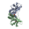
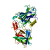
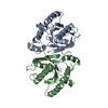
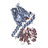
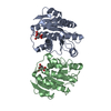
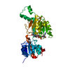
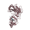
| Entry | Database: PDB / ID: 4emu | ||||||
|---|---|---|---|---|---|---|---|
| Title | Crystal structure of ligand free human STING | ||||||
 Components Components | Transmembrane protein 173 Stimulator of interferon genes Stimulator of interferon genes | ||||||
 Keywords Keywords | MEMBRANE PROTEIN / alpha/beta fold / innate immune sensor / c-di-GMP | ||||||
| Function / homology |  Function and homology information Function and homology informationSTING complex / STAT6-mediated induction of chemokines / protein localization to endoplasmic reticulum / serine/threonine protein kinase complex / 2',3'-cyclic GMP-AMP binding / proton channel activity / cyclic-di-GMP binding / STING mediated induction of host immune responses / IRF3-mediated induction of type I IFN / positive regulation of type I interferon-mediated signaling pathway ...STING complex / STAT6-mediated induction of chemokines / protein localization to endoplasmic reticulum / serine/threonine protein kinase complex / 2',3'-cyclic GMP-AMP binding / proton channel activity / cyclic-di-GMP binding / STING mediated induction of host immune responses / IRF3-mediated induction of type I IFN / positive regulation of type I interferon-mediated signaling pathway / cGAS/STING signaling pathway / reticulophagy / pattern recognition receptor signaling pathway / cytoplasmic pattern recognition receptor signaling pathway / cellular response to exogenous dsRNA / autophagosome membrane / antiviral innate immune response / positive regulation of macroautophagy / autophagosome assembly / cellular response to organic cyclic compound / autophagosome / positive regulation of type I interferon production / cellular response to interferon-beta / signaling adaptor activity / positive regulation of defense response to virus by host / Regulation of innate immune responses to cytosolic DNA / activation of innate immune response / endoplasmic reticulum-Golgi intermediate compartment membrane / positive regulation of interferon-beta production / secretory granule membrane / cytoplasmic vesicle membrane / positive regulation of DNA-binding transcription factor activity / peroxisome / SARS-CoV-1 activates/modulates innate immune responses / positive regulation of protein binding / protein complex oligomerization / regulation of inflammatory response / defense response to virus / RNA polymerase II-specific DNA-binding transcription factor binding / mitochondrial outer membrane / endosome / Golgi membrane / innate immune response / ubiquitin protein ligase binding / Neutrophil degranulation / endoplasmic reticulum membrane / protein kinase binding / SARS-CoV-2 activates/modulates innate and adaptive immune responses / perinuclear region of cytoplasm / protein homodimerization activity / positive regulation of transcription by RNA polymerase II / nucleoplasm / identical protein binding / plasma membrane / cytosolSimilarity search - Function | ||||||
| Biological species |  Homo sapiens (human) Homo sapiens (human) | ||||||
| Method | X-RAY DIFFRACTION / SYNCHROTRON / FOURIER SYNTHESIS / Resolution: 1.9 Å | ||||||
 Authors Authors | Li, P. | ||||||
 Citation Citation | Journal: Nat.Struct.Mol.Biol. / Year: 2012 Title: Structure of STING bound to cyclic di-GMP reveals the mechanism of cyclic dinucleotide recognition by the immune system. Authors: Shu, C. / Yi, G. / Watts, T. / Kao, C.C. / Li, P. | ||||||
| History |
|
- Structure visualization
| Structure viewer | Molecule: MolmilJmol/JSmol |
|---|
- Downloads & links
-Download
| PDBx/mmCIF format | 4emu.cif.gz | 158.6 KB | Display | PDBx/mmCIF format |
|---|---|---|---|---|
| PDB format | pdb4emu.ent.gz | 127.7 KB | Display | PDB format |
| PDBx/mmJSON format | 4emu.json.gz | Tree view | PDBx/mmJSON format | |
| Others |  Other downloads Other downloads |
-Validation report
| Arichive directory | https://data.pdbj.org/pub/pdb/validation_reports/em/4emuftp://data.pdbj.org/pub/pdb/validation_reports/em/4emu | HTTPS FTP |
|---|
-Related structure data


-Links
 PDBj
PDBj


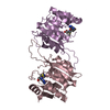
- Assembly
| Deposited unit | 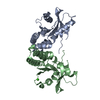
| |||||||||
|---|---|---|---|---|---|---|---|---|---|---|
| 1 |
| |||||||||
| 2 | 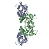
| |||||||||
| Unit cell |
| |||||||||
| Components on special symmetry positions |
|
-Components
| #1: Protein | Stimulator of interferon genes / Endoplasmic reticulum interferon stimulator / ERIS / Mediator of IRF3 activation / hMITA / ...Endoplasmic reticulum interferon stimulator / ERIS / Mediator of IRF3 activation / hMITA / Stimulator of interferon genes protein / hSTING Mass: 21524.158 Da / Num. of mol.: 2 / Fragment: UNP residues 155 - 341 Source method: isolated from a genetically manipulated source Source: (gene. exp.) Homo sapiens (human) / Gene: TMEM173, ERIS, MITA, STING / Production host:  Escherichia coli (E. coli) / References: UniProt: Q86WV6 Escherichia coli (E. coli) / References: UniProt: Q86WV6#2: Chemical | ChemComp-CA / |   Mass: 40.078 Da / Num. of mol.: 1 / Source method: obtained synthetically / Formula: Ca Mass: 40.078 Da / Num. of mol.: 1 / Source method: obtained synthetically / Formula: Ca#3: Water | ChemComp-HOH / | Water Mass: 18.015 Da / Num. of mol.: 146 / Source method: isolated from a natural source / Formula: H2O Mass: 18.015 Da / Num. of mol.: 146 / Source method: isolated from a natural source / Formula: H2O |
|---|
-Experimental details
-Experiment
| Experiment | Method: X-RAY DIFFRACTION / Number of used crystals: 1 |
|---|
- Sample preparation
| Crystal | Density Matthews: 2.03 Å3/Da / Density % sol: 39.3 % |
|---|---|
| Crystal grow | Temperature: 277 K / Method: vapor diffusion, hanging drop / pH: 8.5 Details: 0.1 M HEPES, pH 8.5, 28% PEG400, 0.2M CaCl2, VAPOR DIFFUSION, HANGING DROP, temperature 277K |
-Data collection
| Diffraction | Mean temperature: 120 K |
|---|---|
| Diffraction source | Source: SYNCHROTRON / Site: SSRL  / Beamline: BL11-1 / Wavelength: 0.9794 Å / Beamline: BL11-1 / Wavelength: 0.9794 Å |
| Detector | Type: PSI PILATUS 6M / Detector: PIXEL / Date: Mar 15, 2012 / Details: Si Crystal |
| Radiation | Monochromator: Si Crystal / Protocol: SINGLE WAVELENGTH / Monochromatic (M) / Laue (L): M / Scattering type: x-ray |
| Radiation wavelength | Wavelength: 0.9794 Å / Relative weight: 1 |
| Reflection | Resolution: 1.9→39 Å / Num. all: 27829 / Num. obs: 27829 / % possible obs: 99.6 % / Observed criterion σ(F): 0 / Observed criterion σ(I): 0 |
| Reflection shell | Resolution: 1.9→2 Å / Redundancy: 4.9 % / Rmerge(I) obs: 0.674 / Mean I/σ(I) obs: 2.1 / Rsym value: 0.674 / % possible all: 99.9 |
- Processing
| Software |
| ||||||||||||||||||||||||||||||||||||||||||||||||||||||||||||||||||||||||||||||||||||||||||||||||||||||||||||||||||||||||||||||||||||||||||||||||||||||||||||||||||||||||||||||||||||||||||||||||||||||||||||||||||||||||||||||||||||||||||||||||||||||||||
|---|---|---|---|---|---|---|---|---|---|---|---|---|---|---|---|---|---|---|---|---|---|---|---|---|---|---|---|---|---|---|---|---|---|---|---|---|---|---|---|---|---|---|---|---|---|---|---|---|---|---|---|---|---|---|---|---|---|---|---|---|---|---|---|---|---|---|---|---|---|---|---|---|---|---|---|---|---|---|---|---|---|---|---|---|---|---|---|---|---|---|---|---|---|---|---|---|---|---|---|---|---|---|---|---|---|---|---|---|---|---|---|---|---|---|---|---|---|---|---|---|---|---|---|---|---|---|---|---|---|---|---|---|---|---|---|---|---|---|---|---|---|---|---|---|---|---|---|---|---|---|---|---|---|---|---|---|---|---|---|---|---|---|---|---|---|---|---|---|---|---|---|---|---|---|---|---|---|---|---|---|---|---|---|---|---|---|---|---|---|---|---|---|---|---|---|---|---|---|---|---|---|---|---|---|---|---|---|---|---|---|---|---|---|---|---|---|---|---|---|---|---|---|---|---|---|---|---|---|---|---|---|---|---|---|---|---|---|---|---|---|---|---|---|---|---|---|---|---|---|---|---|
| Refinement | Method to determine structure: FOURIER SYNTHESIS / Resolution: 1.9→37.348 Å / SU ML: 0.57 / σ(F): 1.34 / σ(I): 0 / Phase error: 28.81 / Stereochemistry target values: ML
| ||||||||||||||||||||||||||||||||||||||||||||||||||||||||||||||||||||||||||||||||||||||||||||||||||||||||||||||||||||||||||||||||||||||||||||||||||||||||||||||||||||||||||||||||||||||||||||||||||||||||||||||||||||||||||||||||||||||||||||||||||||||||||
| Solvent computation | Shrinkage radii: 0.83 Å / VDW probe radii: 1.1 Å / Solvent model: FLAT BULK SOLVENT MODEL / Bsol: 45.256 Å2 / ksol: 0.342 e/Å3 | ||||||||||||||||||||||||||||||||||||||||||||||||||||||||||||||||||||||||||||||||||||||||||||||||||||||||||||||||||||||||||||||||||||||||||||||||||||||||||||||||||||||||||||||||||||||||||||||||||||||||||||||||||||||||||||||||||||||||||||||||||||||||||
| Displacement parameters |
| ||||||||||||||||||||||||||||||||||||||||||||||||||||||||||||||||||||||||||||||||||||||||||||||||||||||||||||||||||||||||||||||||||||||||||||||||||||||||||||||||||||||||||||||||||||||||||||||||||||||||||||||||||||||||||||||||||||||||||||||||||||||||||
| Refinement step | Cycle: LAST / Resolution: 1.9→37.348 Å
| ||||||||||||||||||||||||||||||||||||||||||||||||||||||||||||||||||||||||||||||||||||||||||||||||||||||||||||||||||||||||||||||||||||||||||||||||||||||||||||||||||||||||||||||||||||||||||||||||||||||||||||||||||||||||||||||||||||||||||||||||||||||||||
| Refine LS restraints |
| ||||||||||||||||||||||||||||||||||||||||||||||||||||||||||||||||||||||||||||||||||||||||||||||||||||||||||||||||||||||||||||||||||||||||||||||||||||||||||||||||||||||||||||||||||||||||||||||||||||||||||||||||||||||||||||||||||||||||||||||||||||||||||
| LS refinement shell |
| ||||||||||||||||||||||||||||||||||||||||||||||||||||||||||||||||||||||||||||||||||||||||||||||||||||||||||||||||||||||||||||||||||||||||||||||||||||||||||||||||||||||||||||||||||||||||||||||||||||||||||||||||||||||||||||||||||||||||||||||||||||||||||
| Refinement TLS params. | Method: refined / Refine-ID: X-RAY DIFFRACTION
| ||||||||||||||||||||||||||||||||||||||||||||||||||||||||||||||||||||||||||||||||||||||||||||||||||||||||||||||||||||||||||||||||||||||||||||||||||||||||||||||||||||||||||||||||||||||||||||||||||||||||||||||||||||||||||||||||||||||||||||||||||||||||||
| Refinement TLS group |
|
