Monochromator: Side scattering I-beam bent single crystal, asymmetric cut 4.9650 degrees Protocol: SINGLE WAVELENGTH / Monochromatic (M) / Laue (L): M / Scattering type: x-ray
Radiation wavelength
Wavelength: 0.979 Å / Relative weight: 1
Reflection
Resolution: 1.103→92.93 Å / Num. all: 70609 / Num. obs: 70609 / % possible obs: 98.3 % / Redundancy: 3.4 % / Rsym value: 0.089 / Net I/σ(I): 8.3
Reflection shell
Diffraction-ID: 1
Resolution (Å)
Redundancy (%)
Rmerge(I) obs
Mean I/σ(I) obs
Num. measured all
Num. unique all
Rsym value
% possible all
1.103-1.16
3.3
0.483
1.5
34370
10342
0.483
99.9
1.16-1.23
3.4
0.335
2.2
33166
9802
0.335
99.9
1.23-1.32
3.4
0.262
2.8
31661
9277
0.262
99.9
1.32-1.42
3.4
0.194
3.8
29480
8587
0.194
99.8
1.42-1.56
3.4
0.127
5.6
27331
7945
0.127
99.9
1.56-1.74
3.4
0.079
8.9
24667
7235
0.079
99.8
1.74-2.01
3.4
0.068
8.1
21652
6378
0.068
99.6
2.01-2.47
3.2
0.077
7.8
17123
5428
0.077
99.1
2.47-3.49
3.5
0.043
14.2
14361
4155
0.043
96.6
3.49-21.901
2.8
0.052
10.8
4093
1460
0.052
57.9
-
Phasing
Phasing
Method: molecular replacement
Phasing MR
Rfactor: 47.98 / Model details: Phaser MODE: MR_AUTO
Highest resolution
Lowest resolution
Rotation
1.8 Å
21.9 Å
Translation
1.8 Å
21.9 Å
-
Processing
Software
Name
Version
Classification
NB
MOSFLM
3.3.16
datareduction
SCALA
3.3.16
datascaling
PHASER
2.1.4
phasing
REFMAC
refinement
PDB_EXTRACT
3.11
dataextraction
Blu-Ice
datacollection
Refinement
Method to determine structure: MOLECULAR REPLACEMENT / Resolution: 1.103→18.96 Å / Cor.coef. Fo:Fc: 0.967 / Cor.coef. Fo:Fc free: 0.954 / WRfactor Rfree: 0.1778 / WRfactor Rwork: 0.1467 / Occupancy max: 1 / Occupancy min: 0.16 / FOM work R set: 0.9194 / SU B: 0.934 / SU ML: 0.021 / SU R Cruickshank DPI: 0.0319 / SU Rfree: 0.0333 / Cross valid method: THROUGHOUT / σ(F): 0 / ESU R: 0.032 / ESU R Free: 0.033 / Stereochemistry target values: MAXIMUM LIKELIHOOD Details: HYDROGENS HAVE BEEN ADDED IN THE RIDING POSITIONS U VALUES : REFINED INDIVIDUALLY
Rfactor
Num. reflection
% reflection
Selection details
Rfree
0.1762
3543
5 %
RANDOM
Rwork
0.1459
-
-
-
obs
0.1475
70371
97.77 %
-
Solvent computation
Ion probe radii: 0.8 Å / Shrinkage radii: 0.8 Å / VDW probe radii: 1.2 Å / Solvent model: MASK
In the structure databanks used in Yorodumi, some data are registered as the other names, "COVID-19 virus" and "2019-nCoV". Here are the details of the virus and the list of structure data.
Jan 31, 2019. EMDB accession codes are about to change! (news from PDBe EMDB page)
EMDB accession codes are about to change! (news from PDBe EMDB page)
The allocation of 4 digits for EMDB accession codes will soon come to an end. Whilst these codes will remain in use, new EMDB accession codes will include an additional digit and will expand incrementally as the available range of codes is exhausted. The current 4-digit format prefixed with “EMD-” (i.e. EMD-XXXX) will advance to a 5-digit format (i.e. EMD-XXXXX), and so on. It is currently estimated that the 4-digit codes will be depleted around Spring 2019, at which point the 5-digit format will come into force.
The EM Navigator/Yorodumi systems omit the EMD- prefix.
Related info.:Q: What is EMD? / ID/Accession-code notation in Yorodumi/EM Navigator
Yorodumi is a browser for structure data from EMDB, PDB, SASBDB, etc.
This page is also the successor to EM Navigator detail page, and also detail information page/front-end page for Omokage search.
The word "yorodu" (or yorozu) is an old Japanese word meaning "ten thousand". "mi" (miru) is to see.
Related info.:EMDB / PDB / SASBDB / Comparison of 3 databanks / Yorodumi Search / Aug 31, 2016. New EM Navigator & Yorodumi / Yorodumi Papers / Jmol/JSmol / Function and homology information / Changes in new EM Navigator and Yorodumi
 Movie
Movie Controller

 Yorodumi
 Open data
 Basic information
 Components
 Keywords
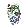
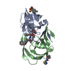
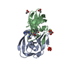
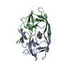
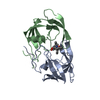
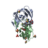
 allostery / fragment binding / HYDROLASE-HYDROLASE INHIBITOR complex
 Function and homology information


 Authors
 Citation
 Structure visualization
 Downloads & links
 Other downloads


 PDBj
PDBj


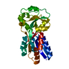
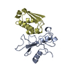
 Assembly
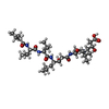
 Type: Oligopeptide


 Mass: 78.133 Da / Num. of mol.: 3 / Source method: obtained synthetically / Formula: C2H6OS
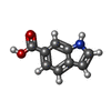
 Mass: 161.157 Da / Num. of mol.: 1 / Source method: obtained synthetically / Formula: C9H7NO2
 Mass: 92.094 Da / Num. of mol.: 1 / Source method: obtained synthetically / Formula: C3H8O3
 Sample preparation
 / Beamline: BL7-1 / Wavelength: 0.979 Å
 Processing