 Movie
Movie Controller


+ Open data

- Basic information
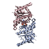
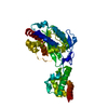
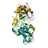
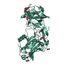
| Entry | Database: PDB / ID: 4ef5 | ||||||
|---|---|---|---|---|---|---|---|
| Title | Crystal structure of STING CTD | ||||||
 Components Components | Transmembrane protein 173 Stimulator of interferon genes Stimulator of interferon genes | ||||||
 Keywords Keywords | IMMUNE SYSTEM / STING/MITA/ERIS/MPYS/TMEM173 / innate immune system / type I interferon / dimerization / c-di-GMP / 5 helices and 5 strands in single domain | ||||||
| Function / homology |  Function and homology information Function and homology informationSTING complex / STAT6-mediated induction of chemokines / protein localization to endoplasmic reticulum / serine/threonine protein kinase complex / 2',3'-cyclic GMP-AMP binding / proton channel activity / cyclic-di-GMP binding / STING mediated induction of host immune responses / IRF3-mediated induction of type I IFN / positive regulation of type I interferon-mediated signaling pathway ...STING complex / STAT6-mediated induction of chemokines / protein localization to endoplasmic reticulum / serine/threonine protein kinase complex / 2',3'-cyclic GMP-AMP binding / proton channel activity / cyclic-di-GMP binding / STING mediated induction of host immune responses / IRF3-mediated induction of type I IFN / positive regulation of type I interferon-mediated signaling pathway / cGAS/STING signaling pathway / reticulophagy / pattern recognition receptor signaling pathway / cellular response to exogenous dsRNA / cytoplasmic pattern recognition receptor signaling pathway / autophagosome membrane / antiviral innate immune response / positive regulation of macroautophagy / autophagosome assembly / cellular response to organic cyclic compound / autophagosome / positive regulation of type I interferon production / cellular response to interferon-beta / signaling adaptor activity / positive regulation of defense response to virus by host / Regulation of innate immune responses to cytosolic DNA / activation of innate immune response / endoplasmic reticulum-Golgi intermediate compartment membrane / positive regulation of interferon-beta production / secretory granule membrane / cytoplasmic vesicle membrane / positive regulation of DNA-binding transcription factor activity / peroxisome / SARS-CoV-1 activates/modulates innate immune responses / positive regulation of protein binding / protein complex oligomerization / regulation of inflammatory response / defense response to virus / RNA polymerase II-specific DNA-binding transcription factor binding / mitochondrial outer membrane / endosome / Golgi membrane / innate immune response / ubiquitin protein ligase binding / Neutrophil degranulation / endoplasmic reticulum membrane / protein kinase binding / SARS-CoV-2 activates/modulates innate and adaptive immune responses / perinuclear region of cytoplasm / protein homodimerization activity / positive regulation of transcription by RNA polymerase II / nucleoplasm / identical protein binding / plasma membrane / cytosolSimilarity search - Function | ||||||
| Biological species |  Homo sapiens (human) Homo sapiens (human) | ||||||
| Method | X-RAY DIFFRACTION / SYNCHROTRON / MOLECULAR REPLACEMENT / Resolution: 2.45 Å | ||||||
 Authors Authors | Ouyang, S. / Ru, H. / Shaw, N. / Jiang, Y. / Niu, F. / Zhu, Y. / Qiu, W. / Li, Y. / Liu, Z.-J. | ||||||
 Citation Citation | Journal: Immunity / Year: 2012 Title: Structural analysis of the STING adaptor protein reveals a hydrophobic dimer interface and mode of cyclic di-GMP binding Authors: Ouyang, S. / Song, X. / Wang, Y. / Ru, H. / Shaw, N. / Jiang, Y. / Niu, F. / Zhu, Y. / Qiu, W. / Parvatiyar, K. / Li, Y. / Zhang, R. / Cheng, G. / Liu, Z.J. | ||||||
| History |
|
- Structure visualization
| Structure viewer | Molecule: MolmilJmol/JSmol |
|---|
- Downloads & links
-Download
| PDBx/mmCIF format | 4ef5.cif.gz | 50.4 KB | Display | PDBx/mmCIF format |
|---|---|---|---|---|
| PDB format | pdb4ef5.ent.gz | 35.7 KB | Display | PDB format |
| PDBx/mmJSON format | 4ef5.json.gz | Tree view | PDBx/mmJSON format | |
| Others |  Other downloads Other downloads |
-Validation report
| Arichive directory | https://data.pdbj.org/pub/pdb/validation_reports/ef/4ef5ftp://data.pdbj.org/pub/pdb/validation_reports/ef/4ef5 | HTTPS FTP |
|---|
-Related structure data


-Links
 PDBj
PDBj


- Assembly
| Deposited unit | 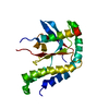
| ||||||||
|---|---|---|---|---|---|---|---|---|---|
| 1 | 
| ||||||||
| Unit cell |
| ||||||||
| Components on special symmetry positions |
|
-Components
| #1: Protein | Stimulator of interferon genes / Endoplasmic reticulum interferon stimulator / ERIS / Mediator of IRF3 activation / hMITA / ...Endoplasmic reticulum interferon stimulator / ERIS / Mediator of IRF3 activation / hMITA / Stimulator of interferon genes protein / hSTING Mass: 29848.463 Da / Num. of mol.: 1 / Fragment: C-TERMINAL DOMAIN Source method: isolated from a genetically manipulated source Source: (gene. exp.) Homo sapiens (human) / Gene: STING/MITA/ERIS/MPYS/TMEM173 / Plasmid: pMCSG7 / Production host:  Escherichia coli (E. coli) / Strain (production host): BL21(DE3) / References: UniProt: Q86WV6 Escherichia coli (E. coli) / Strain (production host): BL21(DE3) / References: UniProt: Q86WV6 |
|---|---|
| #2: Water | ChemComp-HOH / Water Mass: 18.015 Da / Num. of mol.: 21 / Source method: isolated from a natural source / Formula: H2O Mass: 18.015 Da / Num. of mol.: 21 / Source method: isolated from a natural source / Formula: H2O |
-Experimental details
-Experiment
| Experiment | Method: X-RAY DIFFRACTION / Number of used crystals: 1 |
|---|
- Sample preparation
| Crystal | Density Matthews: 2.25 Å3/Da / Density % sol: 45.29 % |
|---|---|
| Crystal grow | Temperature: 289 K / Method: vapor diffusion, hanging drop / pH: 6.5 Details: 14.4%(w/v) PEG8000, 0.08M Cacodylate, 0.16M calcium acetate, 20%(v/v) glycerol, pH 6.5, VAPOR DIFFUSION, HANGING DROP, temperature 289K |
-Data collection
| Diffraction | Mean temperature: 100 K |
|---|---|
| Diffraction source | Source: SYNCHROTRON / Site: SSRF  / Beamline: BL17U / Wavelength: 0.9793 Å / Beamline: BL17U / Wavelength: 0.9793 Å |
| Detector | Type: ADSC QUANTUM 315 / Detector: CCD / Date: Feb 16, 2012 |
| Radiation | Monochromator: GRAPHITE / Protocol: SINGLE WAVELENGTH / Monochromatic (M) / Laue (L): M / Scattering type: x-ray |
| Radiation wavelength | Wavelength: 0.9793 Å / Relative weight: 1 |
| Reflection | Resolution: 2.45→45.18 Å / Num. all: 10182 / Num. obs: 10182 / % possible obs: 99.9 % / Observed criterion σ(F): 2 / Observed criterion σ(I): 2 |
| Reflection shell | Highest resolution: 2.45 Å / % possible all: 99.9 |
- Processing
| Software |
| ||||||||||||||||||||||||
|---|---|---|---|---|---|---|---|---|---|---|---|---|---|---|---|---|---|---|---|---|---|---|---|---|---|
| Refinement | Method to determine structure: MOLECULAR REPLACEMENT / Resolution: 2.45→45.178 Å / Occupancy max: 1 / Occupancy min: 0.44 / FOM work R set: 0.8033 / SU ML: 0.37 / Cross valid method: THROUGHOUT / Phase error: 25.89 / Stereochemistry target values: ML / Details: HYDROGENS HAVE BEEN USED IF PRESENT IN THE INPUT
| ||||||||||||||||||||||||
| Solvent computation | Shrinkage radii: 0.86 Å / VDW probe radii: 1.1 Å / Solvent model: FLAT BULK SOLVENT MODEL / Bsol: 61.567 Å2 / ksol: 0.351 e/Å3 | ||||||||||||||||||||||||
| Displacement parameters | Biso max: 127.43 Å2 / Biso mean: 64.6145 Å2 / Biso min: 28.41 Å2
| ||||||||||||||||||||||||
| Refinement step | Cycle: LAST / Resolution: 2.45→45.178 Å
| ||||||||||||||||||||||||
| Refine LS restraints |
| ||||||||||||||||||||||||
| LS refinement shell | Refine-ID: X-RAY DIFFRACTION / Total num. of bins used: 3 / % reflection obs: 100 %
|
