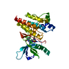
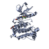
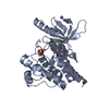
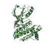
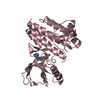
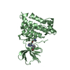
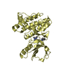
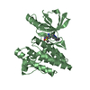
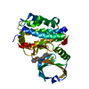
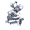
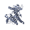
[English] 日本語
Yorodumi
- PDB-4ebw: Structure of Focal Adhesion Kinase catalytic domain in complex wi... -

+
Open data


ID or keywords:

Loading...

-
Basic information

Entry
Database: PDB / ID: 4ebw
TitleStructure of Focal Adhesion Kinase catalytic domain in complex with novel allosteric inhibitor
ComponentsFocal adhesion kinase 1PTK2
KeywordsTRANSFERASE/TRANSFERASE INHIBITOR / Kinase Domain / Allosteric inhibitor / TRANSFERASE-TRANSFERASE INHIBITOR complex
Function / homology
Function and homology information


netrin-activated signaling pathway / regulation of substrate adhesion-dependent cell spreading / regulation of endothelial cell migration / regulation of epithelial cell migration / detection of muscle stretch / positive regulation of ubiquitin-dependent protein catabolic process / JUN kinase binding / signal complex assembly / positive regulation of macrophage proliferation / positive regulation of fibroblast migration ...netrin-activated signaling pathway / regulation of substrate adhesion-dependent cell spreading / regulation of endothelial cell migration / regulation of epithelial cell migration / detection of muscle stretch / positive regulation of ubiquitin-dependent protein catabolic process / JUN kinase binding / signal complex assembly / positive regulation of macrophage proliferation / positive regulation of fibroblast migration / DCC mediated attractive signaling / Signal regulatory protein family interactions / growth hormone receptor signaling pathway / regulation of osteoblast differentiation / MET activates PTK2 signaling / regulation of focal adhesion assembly / establishment of cell polarity / positive regulation of wound healing / positive regulation of macrophage chemotaxis / regulation of GTPase activity / negative regulation of cell-cell adhesion / Fc-gamma receptor signaling pathway involved in phagocytosis / p130Cas linkage to MAPK signaling for integrins / positive regulation of epithelial cell migration / regulation of cytoskeleton organization / Apoptotic cleavage of cellular proteins / regulation of cell adhesion mediated by integrin / GRB2:SOS provides linkage to MAPK signaling for Integrins / negative regulation of anoikis / Estrogen-dependent nuclear events downstream of ESR-membrane signaling / RHO GTPases Activate WASPs and WAVEs / ephrin receptor signaling pathway / positive regulation of protein kinase activity / regulation of cell adhesion / vascular endothelial growth factor receptor signaling pathway / positive regulation of epithelial to mesenchymal transition / heart morphogenesis / stress fiber / EPHB-mediated forward signaling / NCAM signaling for neurite out-growth / SH2 domain binding / Integrin signaling / transforming growth factor beta receptor signaling pathway / ciliary basal body / protein tyrosine phosphatase activity / molecular function activator activity / integrin-mediated signaling pathway / cell motility / placenta development / FCGR3A-mediated phagocytosis / axon guidance / regulation of protein phosphorylation / non-specific protein-tyrosine kinase / non-membrane spanning protein tyrosine kinase activity / epidermal growth factor receptor signaling pathway / Regulation of actin dynamics for phagocytic cup formation / VEGFA-VEGFR2 Pathway / peptidyl-tyrosine phosphorylation / cell migration / integrin binding / actin binding / regulation of cell population proliferation / cell cortex / regulation of cell shape / RAF/MAP kinase cascade / protein phosphatase binding / protein tyrosine kinase activity / angiogenesis / dendritic spine / protein autophosphorylation / Extra-nuclear estrogen signaling / positive regulation of phosphatidylinositol 3-kinase/protein kinase B signal transduction / cytoskeleton / positive regulation of cell migration / positive regulation of protein phosphorylation / intracellular membrane-bounded organelle / focal adhesion / centrosome / positive regulation of cell population proliferation / negative regulation of apoptotic process / protein kinase binding / perinuclear region of cytoplasm / ATP binding / nucleus / plasma membrane / cytosol / cytoplasm
Similarity search - Function
Focal adhesion kinase, targeting (FAT) domain / Focal adhesion kinase, targeting (FAT) domain superfamily / Focal adhesion kinase, N-terminal / FAK1/PYK2, FERM domain C-lobe / Focal adhesion targeting region / FERM N-terminal domain / : / FAK1/PYK2, FERM domain C-lobe / FERM domain signature 2. / FERM central domain ...Focal adhesion kinase, targeting (FAT) domain / Focal adhesion kinase, targeting (FAT) domain superfamily / Focal adhesion kinase, N-terminal / FAK1/PYK2, FERM domain C-lobe / Focal adhesion targeting region / FERM N-terminal domain / : / FAK1/PYK2, FERM domain C-lobe / FERM domain signature 2. / FERM central domain / FERM/acyl-CoA-binding protein superfamily / FERM central domain / FERM superfamily, second domain / FERM domain / FERM domain profile. / Band 4.1 domain / Band 4.1 homologues / Tyrosine-protein kinase, catalytic domain / Tyrosine kinase, catalytic domain / Tyrosine protein kinases specific active-site signature. / PH-like domain superfamily / Tyrosine-protein kinase, active site / Protein tyrosine and serine/threonine kinase / Serine-threonine/tyrosine-protein kinase, catalytic domain / Ubiquitin-like domain superfamily / Transferase(Phosphotransferase) domain 1 / Transferase(Phosphotransferase); domain 1 / Phosphorylase Kinase; domain 1 / Phosphorylase Kinase; domain 1 / Protein kinase, ATP binding site / Protein kinases ATP-binding region signature. / Protein kinase domain profile. / Protein kinase domain / Protein kinase-like domain superfamily / 2-Layer Sandwich / Orthogonal Bundle / Mainly Alpha / Alpha Beta
Similarity search - Domain/homology
Chem-0PF / Focal adhesion kinase 1
Similarity search - Component
Biological speciesHomo sapiens (human)
MethodX-RAY DIFFRACTION / SYNCHROTRON / MOLECULAR REPLACEMENT / Resolution: 2.65 Å
AuthorsIwatani, M. / Iwata, H. / Okabe, A. / Skene, R.J. / Tomita, N. / Hayashi, Y. / Aramaki, Y. / Hosfield, D.J. / Hori, A. / Baba, A. / Miki, H.
CitationJournal: Eur.J.Med.Chem. / Year: 2013
Title: Discovery and characterization of novel allosteric FAK inhibitors.
Authors: Iwatani, M. / Iwata, H. / Okabe, A. / Skene, R.J. / Tomita, N. / Hayashi, Y. / Aramaki, Y. / Hosfield, D.J. / Hori, A. / Baba, A. / Miki, H.
History
DepositionMar 25, 2012Deposition site: RCSB / Processing site: RCSB
Revision 1.0Jul 25, 2012Provider: repository / Type: Initial release
Revision 1.1Jan 2, 2013Group: Database references
Revision 1.2Jan 16, 2013Group: Database references
Revision 1.3Mar 27, 2013Group: Database references

-
Structure visualization

Structure viewerMolecule:
MolmilJmol/JSmol

Downloads & links

-
Assembly

Deposited unit
A: Focal adhesion kinase 1
hetero molecules


Theoretical massNumber of molelcules
Total (without water)35,4622
Polymers35,0941
Non-polymers3671
Water86548
1


  • Idetical with deposited unit
  • defined by author&software
TypeNameSymmetry operationNumber
identity operation1_555x,y,z1
MethodPISA
Unit cell
Length a, b, c (Å)81.300, 47.860, 83.667
Angle α, β, γ (deg.)90.00, 118.39, 90.00
Int Tables number5
Space group name H-MC121

-
Components

#1: Protein Focal adhesion kinase 1 / PTK2 / FADK 1 / Focal adhesion kinase-related nonkinase / FRNK / Protein phosphatase 1 regulatory subunit ...FADK 1 / Focal adhesion kinase-related nonkinase / FRNK / Protein phosphatase 1 regulatory subunit 71 / PPP1R71 / Protein-tyrosine kinase 2 / p125FAK / pp125FAK


Mass: 35094.312 Da / Num. of mol.: 1 / Fragment: unp residues 411-686
Source method: isolated from a genetically manipulated source
Source: (gene. exp.) Homo sapiens (human) / Gene: PTK2, FAK, FAK1 / Production host: Spodoptera frugiperda (fall armyworm)
References: UniProt: Q05397, non-specific protein-tyrosine kinase
#2: Chemical ChemComp-0PF / 1-ethyl-8-(4-ethylphenyl)-5-methyl-1,5-dihydropyrazolo[4,3-c][2,1]benzothiazine 4,4-dioxide


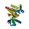
Mass: 367.465 Da / Num. of mol.: 1 / Source method: obtained synthetically / Formula: C20H21N3O2S
#3: Water ChemComp-HOH / water / Water


Mass: 18.015 Da / Num. of mol.: 48 / Source method: isolated from a natural source / Formula: H2O

-
Experimental details

-
Experiment

ExperimentMethod: X-RAY DIFFRACTION

-
Sample preparation

CrystalDensity Matthews: 2.04 Å3/Da / Density % sol: 39.71 %
Crystal growTemperature: 293 K / Method: vapor diffusion, sitting drop / pH: 7.5
Details: 20% PEG4000, 8% 2-Propanol, 12% Glycerol, 0.1M HEPES, pH 7.5, VAPOR DIFFUSION, SITTING DROP, temperature 293K

-
Data collection

Diffraction sourceSource: SYNCHROTRON / Site: SSRL / Beamline: BL7-1 / Wavelength: 0.97 Å
DetectorType: ADSC QUANTUM 315r / Detector: CCD
RadiationProtocol: SINGLE WAVELENGTH / Monochromatic (M) / Laue (L): M / Scattering type: x-ray
Radiation wavelengthWavelength: 0.97 Å / Relative weight: 1
ReflectionResolution: 2.6→50 Å / Num. all: 15207 / Num. obs: 8832 / % possible obs: 91.5 %

-
Processing

Software
NameVersionClassification
HKL-2000data collection
PHASERphasing
REFMAC5.5.0109refinement
HKL-2000data reduction
HKL-2000data scaling
RefinementMethod to determine structure: MOLECULAR REPLACEMENT / Resolution: 2.65→30 Å / Cor.coef. Fo:Fc: 0.95 / Cor.coef. Fo:Fc free: 0.878 / SU B: 30.501 / SU ML: 0.296 / Cross valid method: THROUGHOUT / ESU R Free: 0.41 / Stereochemistry target values: MAXIMUM LIKELIHOOD
RfactorNum. reflection% reflectionSelection details
Rfree0.27349 373 4.8 %RANDOM
Rwork0.18209 ---
all0.18629 15207 --
obs0.18629 7418 92.86 %-
Solvent computationIon probe radii: 0.8 Å / Shrinkage radii: 0.8 Å / VDW probe radii: 1.4 Å / Solvent model: MASK
Displacement parametersBiso mean: 52.106 Å2
Baniso -1Baniso -2Baniso -3
1-7.62 Å20 Å21.39 Å2
2---2.74 Å20 Å2
3----3.56 Å2
Refinement stepCycle: LAST / Resolution: 2.65→30 Å
ProteinNucleic acidLigandSolventTotal
Num. atoms2081 0 26 48 2155
Refine LS restraints
Refine-IDTypeDev idealDev ideal targetNumber
X-RAY DIFFRACTIONr_bond_refined_d0.0110.0222160
X-RAY DIFFRACTIONr_angle_refined_deg1.2991.982924
X-RAY DIFFRACTIONr_angle_other_deg
X-RAY DIFFRACTIONr_dihedral_angle_1_deg5.2765256
X-RAY DIFFRACTIONr_dihedral_angle_2_deg33.30823.22996
X-RAY DIFFRACTIONr_dihedral_angle_3_deg15.17515388
X-RAY DIFFRACTIONr_dihedral_angle_4_deg15.4931518
X-RAY DIFFRACTIONr_chiral_restr0.0890.2321
X-RAY DIFFRACTIONr_gen_planes_refined0.0050.0211612
X-RAY DIFFRACTIONr_gen_planes_other
X-RAY DIFFRACTIONr_nbd_refined
X-RAY DIFFRACTIONr_nbd_other
X-RAY DIFFRACTIONr_nbtor_refined
X-RAY DIFFRACTIONr_nbtor_other
X-RAY DIFFRACTIONr_xyhbond_nbd_refined
X-RAY DIFFRACTIONr_xyhbond_nbd_other
X-RAY DIFFRACTIONr_metal_ion_refined
X-RAY DIFFRACTIONr_metal_ion_other
X-RAY DIFFRACTIONr_symmetry_vdw_refined
X-RAY DIFFRACTIONr_symmetry_vdw_other
X-RAY DIFFRACTIONr_symmetry_hbond_refined
X-RAY DIFFRACTIONr_symmetry_hbond_other
X-RAY DIFFRACTIONr_symmetry_metal_ion_refined
X-RAY DIFFRACTIONr_symmetry_metal_ion_other
X-RAY DIFFRACTIONr_mcbond_it0.6931.51293
X-RAY DIFFRACTIONr_mcbond_other
X-RAY DIFFRACTIONr_mcangle_it1.33322098
X-RAY DIFFRACTIONr_scbond_it2.2383867
X-RAY DIFFRACTIONr_scangle_it3.644.5826
X-RAY DIFFRACTIONr_rigid_bond_restr
X-RAY DIFFRACTIONr_sphericity_free
X-RAY DIFFRACTIONr_sphericity_bonded
LS refinement shellResolution: 2.65→2.718 Å / Total num. of bins used: 20
RfactorNum. reflection% reflection
Rfree0.369 19 -
Rwork0.228 410 -
obs--70.56 %
Refinement TLS params.

Method: refined / Refine-ID: X-RAY DIFFRACTION

IDL112)L122)L132)L222)L232)L332)S11 (Å °)S12 (Å °)S13 (Å °)S21 (Å °)S22 (Å °)S23 (Å °)S31 (Å °)S32 (Å °)S33 (Å °)T112)T122)T132)T222)T232)T332)Origin x (Å)Origin y (Å)Origin z (Å)
14.78641.84010.88728.5877-0.86226.71840.03020.29020.1002-0.4753-0.0108-0.13080.01530.2202-0.01940.0573-0.0024-0.04740.18890.05840.153214.983212.31782.9056
21.26640.57520.04314.6864-0.02414.63580.02140.02580.18660.16750.13570.3953-0.3265-0.1365-0.15710.0396-0.02270.00670.28180.05320.258311.590113.726210.8643
31.3338-0.7194-1.26785.69551.97774.7955-0.1033-0.0594-0.05090.61110.16730.16980.21370.2286-0.0640.13870.021-0.01540.17650.02150.167113.1654-3.138319.8951
42.89670.0505-2.33544.63712.29535.6292-0.19970.26410.00350.6387-0.12350.51080.3338-0.47870.32310.1978-0.00670.07880.17980.00830.17723.8511.156724.3881
56.2578-1.1609-2.25174.77722.79595.764-0.35360.3133-0.31511.0456-0.24910.89361.1044-0.58330.60270.5287-0.13380.26130.1438-0.0420.23451.9773-11.319127.2665
611.6979-1.676-7.48096.16493.93628.0642-0.7039-1.2104-0.46332.46490.3293-0.18891.05411.18690.37461.17640.1067-0.09990.30280.12180.091614.4086-7.658133.5795
Refinement TLS group
IDRefine-IDRefine TLS-IDAuth asym-IDAuth seq-ID
1X-RAY DIFFRACTION1A411 - 455
2X-RAY DIFFRACTION2A456 - 505
3X-RAY DIFFRACTION3A506 - 555
4X-RAY DIFFRACTION4A556 - 605
5X-RAY DIFFRACTION5A606 - 655
6X-RAY DIFFRACTION6A656 - 686

+
About Yorodumi

-
News

-
Feb 9, 2022. New format data for meta-information of EMDB entries

New format data for meta-information of EMDB entries

  • Version 3 of the EMDB header file is now the official format.
  • The previous official version 1.9 will be removed from the archive.

Related info.:EMDB header

External links:wwPDB to switch to version 3 of the EMDB data model

-
Aug 12, 2020. Covid-19 info

Covid-19 info

URL: https://pdbj.org/emnavi/covid19.php

New page: Covid-19 featured information page in EM Navigator.

Related info.:Covid-19 info / Mar 5, 2020. Novel coronavirus structure data

+
Mar 5, 2020. Novel coronavirus structure data

Novel coronavirus structure data

Related info.:Yorodumi Speices / Aug 12, 2020. Covid-19 info

External links:COVID-19 featured content - PDBj / Molecule of the Month (242):Coronavirus Proteases

+
Jan 31, 2019. EMDB accession codes are about to change! (news from PDBe EMDB page)

EMDB accession codes are about to change! (news from PDBe EMDB page)

  • The allocation of 4 digits for EMDB accession codes will soon come to an end. Whilst these codes will remain in use, new EMDB accession codes will include an additional digit and will expand incrementally as the available range of codes is exhausted. The current 4-digit format prefixed with “EMD-” (i.e. EMD-XXXX) will advance to a 5-digit format (i.e. EMD-XXXXX), and so on. It is currently estimated that the 4-digit codes will be depleted around Spring 2019, at which point the 5-digit format will come into force.
  • The EM Navigator/Yorodumi systems omit the EMD- prefix.

Related info.:Q: What is EMD? / ID/Accession-code notation in Yorodumi/EM Navigator

External links:EMDB Accession Codes are Changing Soon! / Contact to PDBj

+
Jul 12, 2017. Major update of PDB

Major update of PDB

  • wwPDB released updated PDB data conforming to the new PDBx/mmCIF dictionary.
  • This is a major update changing the version number from 4 to 5, and with Remediation, in which all the entries are updated.
  • In this update, many items about electron microscopy experimental information are reorganized (e.g. em_software).
  • Now, EM Navigator and Yorodumi are based on the updated data.

External links:wwPDB Remediation / Enriched Model Files Conforming to OneDep Data Standards Now Available in the PDB FTP Archive

-
Yorodumi

Thousand views of thousand structures

  • Yorodumi is a browser for structure data from EMDB, PDB, SASBDB, etc.
  • This page is also the successor to EM Navigator detail page, and also detail information page/front-end page for Omokage search.
  • The word "yorodu" (or yorozu) is an old Japanese word meaning "ten thousand". "mi" (miru) is to see.

Related info.:EMDB / PDB / SASBDB / Comparison of 3 databanks / Yorodumi Search / Aug 31, 2016. New EM Navigator & Yorodumi / Yorodumi Papers / Jmol/JSmol / Function and homology information / Changes in new EM Navigator and Yorodumi

Read more