 Movie
Movie Controller

[English] 日本語
 Yorodumi
- PDB-4e5o: Crystal structure of mouse thymidylate synthase in complex with dUMP -
+ Open data

- Basic information
| Entry | Database: PDB / ID: 4e5o | |||||||||
|---|---|---|---|---|---|---|---|---|---|---|
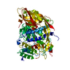
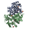
| Title | Crystal structure of mouse thymidylate synthase in complex with dUMP | |||||||||
 Components Components | Thymidylate synthase | |||||||||
 Keywords Keywords | TRANSFERASE / protein-ligand complex / methyltransferase / Nucleotide biosynthesis | |||||||||
| Function / homology |  Function and homology information Function and homology informationInterconversion of nucleotide di- and triphosphates / uracil metabolic process / response to organophosphorus / intestinal epithelial cell maturation / response to folic acid / response to vitamin A / thymidylate synthase / sequence-specific mRNA binding / cartilage development / tetrahydrofolate interconversion ...Interconversion of nucleotide di- and triphosphates / uracil metabolic process / response to organophosphorus / intestinal epithelial cell maturation / response to folic acid / response to vitamin A / thymidylate synthase / sequence-specific mRNA binding / cartilage development / tetrahydrofolate interconversion / thymidylate synthase activity / folic acid binding / dTMP biosynthetic process / dTTP biosynthetic process / heterocyclic compound binding / developmental growth / response to glucocorticoid / mRNA regulatory element binding translation repressor activity / response to progesterone / response to cytokine / liver regeneration / response to toxic substance / circadian rhythm / regulation of translation / methylation / response to ethanol / mitochondrial inner membrane / negative regulation of translation / mitochondrial matrix / response to xenobiotic stimulus / mRNA binding / protein homodimerization activity / mitochondrion / nucleus / cytosol / cytoplasmSimilarity search - Function | |||||||||
| Biological species |  Mus musculus (house mouse) Mus musculus (house mouse) | |||||||||
| Method | X-RAY DIFFRACTION / SYNCHROTRON / MOLECULAR REPLACEMENT / molecular replacement / Resolution: 1.7 Å | |||||||||
 Authors Authors | Dowiercial, A. / Jarmula, A. / Rypniewski, W. / Sokolowska, M. / Fraczyk, T. / Ciesla, J. / Rode, W. | |||||||||
 Citation Citation | Journal: Struct Chem / Year: 2016 Title: Mouse thymidylate synthase does not show the inactive conformation, observed for the human enzyme Authors: Dowiercial, A. / Jarmula, A. / Wilk, P. / Rypniewski, W. / Kowalska, M. / Fraczyk, T. / Ciesla, J. / Rode, W. #1: Journal: Pteridines / Year: 2009Title: Crystal structures of substrate- and sulfate-bound mouse thymidylate synthase Authors: Dowiercial, A. / Jarmula, A. / Rypniewski, W. / Sokolowska, M. / Fraczyk, T. / Ciesla, J. / Rode, W. | |||||||||
| History |
|
- Structure visualization
| Structure viewer | Molecule: MolmilJmol/JSmol |
|---|
- Downloads & links
-Download
| PDBx/mmCIF format | 4e5o.cif.gz | 384.4 KB | Display | PDBx/mmCIF format |
|---|---|---|---|---|
| PDB format | pdb4e5o.ent.gz | 311.1 KB | Display | PDB format |
| PDBx/mmJSON format | 4e5o.json.gz | Tree view | PDBx/mmJSON format | |
| Others |  Other downloads Other downloads |
-Validation report
| Arichive directory | https://data.pdbj.org/pub/pdb/validation_reports/e5/4e5oftp://data.pdbj.org/pub/pdb/validation_reports/e5/4e5o | HTTPS FTP |
|---|
-Related structure data
| Related structure data | 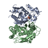 3ihiC  5fctC 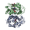 1rtsS C: citing same article ( S: Starting model for refinement |
|---|---|
| Similar structure data |


-Links
 PDBj
PDBj


- Assembly
| Deposited unit | 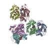
| ||||||||
|---|---|---|---|---|---|---|---|---|---|
| 1 | 
| ||||||||
| 2 | 
| ||||||||
| 3 | 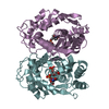
| ||||||||
| Unit cell |
| ||||||||
| Components on special symmetry positions |
|
-Components
| #1: Protein | / TS / TSase Mass: 35001.016 Da / Num. of mol.: 6 Source method: isolated from a genetically manipulated source Source: (gene. exp.) Mus musculus (house mouse) / Gene: Tyms / Plasmid: pPIGDM4 / Production host:  Escherichia coli (E. coli) / Strain (production host): TX61- / References: UniProt: P07607, thymidylate synthase Escherichia coli (E. coli) / Strain (production host): TX61- / References: UniProt: P07607, thymidylate synthase#2: Chemical | ChemComp-UMP / Deoxyuridine monophosphate  Mass: 308.182 Da / Num. of mol.: 7 / Source method: obtained synthetically / Formula: C9H13N2O8P Mass: 308.182 Da / Num. of mol.: 7 / Source method: obtained synthetically / Formula: C9H13N2O8P#3: Chemical | 1,4-Butanediol  Mass: 90.121 Da / Num. of mol.: 3 / Source method: obtained synthetically / Formula: C4H10O2 Mass: 90.121 Da / Num. of mol.: 3 / Source method: obtained synthetically / Formula: C4H10O2#4: Water | ChemComp-HOH / | Water Mass: 18.015 Da / Num. of mol.: 1472 / Source method: isolated from a natural source / Formula: H2O Mass: 18.015 Da / Num. of mol.: 1472 / Source method: isolated from a natural source / Formula: H2O |
|---|
-Experimental details
-Experiment
| Experiment | Method: X-RAY DIFFRACTION / Number of used crystals: 1 |
|---|
- Sample preparation
| Crystal | Density Matthews: 2.3 Å3/Da / Density % sol: 46.49 % |
|---|---|
| Crystal grow | Temperature: 278 K / Method: vapor diffusion, hanging drop Details: K/Na Tartrate, PEG 3350, VAPOR DIFFUSION, HANGING DROP, temperature 278K |
-Data collection
| Diffraction | Mean temperature: 100 K | ||||||||||||||||||||||||||||||||||||||||||||||||||||||||||||||||||||||||||||||||||||||||||||||||||||||||||||||||||||||||||||||
|---|---|---|---|---|---|---|---|---|---|---|---|---|---|---|---|---|---|---|---|---|---|---|---|---|---|---|---|---|---|---|---|---|---|---|---|---|---|---|---|---|---|---|---|---|---|---|---|---|---|---|---|---|---|---|---|---|---|---|---|---|---|---|---|---|---|---|---|---|---|---|---|---|---|---|---|---|---|---|---|---|---|---|---|---|---|---|---|---|---|---|---|---|---|---|---|---|---|---|---|---|---|---|---|---|---|---|---|---|---|---|---|---|---|---|---|---|---|---|---|---|---|---|---|---|---|---|---|
| Diffraction source | Source: SYNCHROTRON / Site: MAX II  / Beamline: I911-5 / Wavelength: 0.908 Å / Beamline: I911-5 / Wavelength: 0.908 Å | ||||||||||||||||||||||||||||||||||||||||||||||||||||||||||||||||||||||||||||||||||||||||||||||||||||||||||||||||||||||||||||||
| Detector | Type: MAR CCD 165 mm / Detector: CCD / Date: May 8, 2008 | ||||||||||||||||||||||||||||||||||||||||||||||||||||||||||||||||||||||||||||||||||||||||||||||||||||||||||||||||||||||||||||||
| Radiation | Monochromator: Si(111) bent crystal, horizontally focusing / Protocol: SINGLE WAVELENGTH / Monochromatic (M) / Laue (L): M / Scattering type: x-ray | ||||||||||||||||||||||||||||||||||||||||||||||||||||||||||||||||||||||||||||||||||||||||||||||||||||||||||||||||||||||||||||||
| Radiation wavelength | Wavelength: 0.908 Å / Relative weight: 1 | ||||||||||||||||||||||||||||||||||||||||||||||||||||||||||||||||||||||||||||||||||||||||||||||||||||||||||||||||||||||||||||||
| Reflection | Resolution: 1.7→20 Å / Num. obs: 207806 / % possible obs: 99.9 % / Rmerge(I) obs: 0.063 / Χ2: 0.955 / Net I/σ(I): 16.9 | ||||||||||||||||||||||||||||||||||||||||||||||||||||||||||||||||||||||||||||||||||||||||||||||||||||||||||||||||||||||||||||||
| Reflection shell |
|
-Phasing
| Phasing | Method: molecular replacement | |||||||||
|---|---|---|---|---|---|---|---|---|---|---|
| Phasing MR | Model details: Phaser MODE: MR_AUTO
|
- Processing
| Software |
| |||||||||||||||||||||||||||||||||||||||||||||||||||||||||||||||||
|---|---|---|---|---|---|---|---|---|---|---|---|---|---|---|---|---|---|---|---|---|---|---|---|---|---|---|---|---|---|---|---|---|---|---|---|---|---|---|---|---|---|---|---|---|---|---|---|---|---|---|---|---|---|---|---|---|---|---|---|---|---|---|---|---|---|---|
| Refinement | Method to determine structure: MOLECULAR REPLACEMENT Starting model: PDB ENTRY 1RTS Resolution: 1.7→19.95 Å / Cor.coef. Fo:Fc: 0.938 / Cor.coef. Fo:Fc free: 0.9 / WRfactor Rfree: 0.3058 / WRfactor Rwork: 0.2486 / Occupancy max: 1 / Occupancy min: 0.3 / FOM work R set: 0.7769 / SU B: 2.855 / SU ML: 0.095 / SU R Cruickshank DPI: 0.1348 / SU Rfree: 0.1395 / Cross valid method: THROUGHOUT / σ(F): 0 / ESU R: 0.135 / ESU R Free: 0.139 / Stereochemistry target values: MAXIMUM LIKELIHOOD
| |||||||||||||||||||||||||||||||||||||||||||||||||||||||||||||||||
| Solvent computation | Ion probe radii: 0.8 Å / Shrinkage radii: 0.8 Å / VDW probe radii: 1.4 Å / Solvent model: MASK | |||||||||||||||||||||||||||||||||||||||||||||||||||||||||||||||||
| Displacement parameters | Biso max: 87.12 Å2 / Biso mean: 29.3331 Å2 / Biso min: 9.21 Å2
| |||||||||||||||||||||||||||||||||||||||||||||||||||||||||||||||||
| Refinement step | Cycle: LAST / Resolution: 1.7→19.95 Å
| |||||||||||||||||||||||||||||||||||||||||||||||||||||||||||||||||
| Refine LS restraints |
| |||||||||||||||||||||||||||||||||||||||||||||||||||||||||||||||||
| LS refinement shell | Resolution: 1.7→1.745 Å / Total num. of bins used: 20
|
