 Movie
Movie Controller


+ Open data

- Basic information
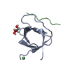
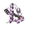
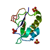
| Entry | Database: PDB / ID: 4e1i | ||||||
|---|---|---|---|---|---|---|---|
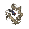
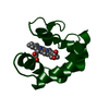
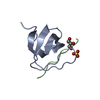
| Title | Fragment of human prion protein | ||||||
 Components Components | (Major prion protein) x 2 | ||||||
 Keywords Keywords |  CELL CYCLE / beta prism / amyloid-related oligomer / PROTEIN FIBRIL CELL CYCLE / beta prism / amyloid-related oligomer / PROTEIN FIBRIL | ||||||
| Function / homology |  Function and homology information Function and homology informationpositive regulation of glutamate receptor signaling pathway / negative regulation of amyloid precursor protein catabolic process / lamin binding / regulation of glutamate receptor signaling pathway / regulation of calcium ion import across plasma membrane / aspartic-type endopeptidase inhibitor activity / glycosaminoglycan binding / ATP-dependent protein binding / regulation of potassium ion transmembrane transport / NCAM1 interactions ...positive regulation of glutamate receptor signaling pathway / negative regulation of amyloid precursor protein catabolic process / lamin binding / regulation of glutamate receptor signaling pathway / regulation of calcium ion import across plasma membrane / aspartic-type endopeptidase inhibitor activity / glycosaminoglycan binding / ATP-dependent protein binding / regulation of potassium ion transmembrane transport / NCAM1 interactions / negative regulation of interleukin-17 production / negative regulation of dendritic spine maintenance / type 5 metabotropic glutamate receptor binding / cupric ion binding / negative regulation of protein processing / negative regulation of calcineurin-NFAT signaling cascade / dendritic spine maintenance / negative regulation of interleukin-2 production / negative regulation of T cell receptor signaling pathway / Insertion of tail-anchored proteins into the endoplasmic reticulum membrane / extrinsic component of membrane / cuprous ion binding / negative regulation of amyloid-beta formation / negative regulation of activated T cell proliferation / response to amyloid-beta / : / negative regulation of type II interferon production / intracellular copper ion homeostasis / negative regulation of long-term synaptic potentiation / positive regulation of protein targeting to membrane / long-term memory / response to cadmium ion / regulation of peptidyl-tyrosine phosphorylation / inclusion body / cellular response to copper ion / neuron projection maintenance / tubulin binding / negative regulation of protein phosphorylation / molecular condensate scaffold activity / molecular function activator activity / positive regulation of protein localization to plasma membrane / protein destabilization / protein homooligomerization / negative regulation of DNA-binding transcription factor activity / terminal bouton / cellular response to amyloid-beta / positive regulation of peptidyl-tyrosine phosphorylation / positive regulation of neuron apoptotic process / cellular response to xenobiotic stimulus / signaling receptor activity / amyloid-beta binding / protein-folding chaperone binding / postsynapse / microtubule binding / nuclear membrane / protease binding / response to oxidative stress / transmembrane transporter binding / postsynaptic density / molecular adaptor activity / learning or memory / regulation of cell cycle / membrane raft / copper ion binding / cell cycle / external side of plasma membrane / intracellular membrane-bounded organelle / dendrite / protein-containing complex binding / negative regulation of apoptotic process / Golgi apparatus / cell surface / endoplasmic reticulum / extracellular exosome / identical protein binding / plasma membrane / cytosol / cytoplasmSimilarity search - Function | ||||||
| Biological species |  Homo sapiens (human) Homo sapiens (human) | ||||||
| Method | X-RAY DIFFRACTION / molecular replacement / Resolution: 2.03 Å | ||||||
 Authors Authors | Apostol, M.I. / Surewicz, W.K. | ||||||
 Citation Citation | Journal: J.Am.Chem.Soc. / Year: 2013 Title: Crystal structure of a human prion protein fragment reveals a motif for oligomer formation. Authors: Apostol, M.I. / Perry, K. / Surewicz, W.K. | ||||||
| History |
|
- Structure visualization
| Structure viewer | Molecule: MolmilJmol/JSmol |
|---|
- Downloads & links
-Download
| PDBx/mmCIF format | 4e1i.cif.gz | 27.3 KB | Display | PDBx/mmCIF format |
|---|---|---|---|---|
| PDB format | pdb4e1i.ent.gz | 21 KB | Display | PDB format |
| PDBx/mmJSON format | 4e1i.json.gz | Tree view | PDBx/mmJSON format | |
| Others |  Other downloads Other downloads |
-Validation report
| Arichive directory | https://data.pdbj.org/pub/pdb/validation_reports/e1/4e1iftp://data.pdbj.org/pub/pdb/validation_reports/e1/4e1i | HTTPS FTP |
|---|
-Related structure data


-Links
 PDBj
PDBj


- Assembly
| Deposited unit | 
| ||||||||
|---|---|---|---|---|---|---|---|---|---|
| 1 |
| ||||||||
| Unit cell |
|
-Components
| #1: Protein/peptide | Mass: 700.784 Da / Num. of mol.: 6 / Fragment: UNP residues 177-182 / Source method: obtained synthetically / Source: (synth.) Homo sapiens (human) / References: UniProt: P04156#2: Protein/peptide | Mass: 723.858 Da / Num. of mol.: 6 / Fragment: UNP residues 211-216 / Source method: obtained synthetically / Source: (synth.) Homo sapiens (human) / References: UniProt: P04156#3: Water | ChemComp-HOH / | Water Mass: 18.015 Da / Num. of mol.: 67 / Source method: isolated from a natural source / Formula: H2O Mass: 18.015 Da / Num. of mol.: 67 / Source method: isolated from a natural source / Formula: H2OCompound details | FRAGMENT OF HUMAN PRION PROTEIN FROM HELIX 2 AND 3 | Source details | SEGMENTS CORRESPONDING TO RESIDUES 177-182 AND 211-216 OF HUMAN PRION WAS LINKED BY DISULFIDE BOND ...SEGMENTS CORRESPOND | |
|---|
-Experimental details
-Experiment
| Experiment | Method: X-RAY DIFFRACTION / Number of used crystals: 1 |
|---|
- Sample preparation
| Crystal | Density Matthews: 1.99 Å3/Da / Density % sol: 38.04 % |
|---|---|
| Crystal grow | Temperature: 298 K / Method: vapor diffusion, hanging drop / pH: 5.5 Details: 100mM Bis Tris pH 5.5, 25% PEG 3350, vapor diffusion, hanging drop, temperature 298K |
-Data collection
| Diffraction | Mean temperature: 100 K | ||||||||||||||||||||||||||||||||||||||||||||||||||||||||||||||||||||||||||||||||||||||||
|---|---|---|---|---|---|---|---|---|---|---|---|---|---|---|---|---|---|---|---|---|---|---|---|---|---|---|---|---|---|---|---|---|---|---|---|---|---|---|---|---|---|---|---|---|---|---|---|---|---|---|---|---|---|---|---|---|---|---|---|---|---|---|---|---|---|---|---|---|---|---|---|---|---|---|---|---|---|---|---|---|---|---|---|---|---|---|---|---|---|
| Diffraction source | Source: ROTATING ANODE / Type: RIGAKU MICROMAX-007 HF / Wavelength: 1.5418 Å | ||||||||||||||||||||||||||||||||||||||||||||||||||||||||||||||||||||||||||||||||||||||||
| Detector | Type: RIGAKU SATURN 944+ / Detector: CCD / Date: Oct 13, 2010 | ||||||||||||||||||||||||||||||||||||||||||||||||||||||||||||||||||||||||||||||||||||||||
| Radiation | Protocol: SINGLE WAVELENGTH / Monochromatic (M) / Laue (L): M / Scattering type: x-ray | ||||||||||||||||||||||||||||||||||||||||||||||||||||||||||||||||||||||||||||||||||||||||
| Radiation wavelength | Wavelength: 1.5418 Å / Relative weight: 1 | ||||||||||||||||||||||||||||||||||||||||||||||||||||||||||||||||||||||||||||||||||||||||
| Reflection | Resolution: 2.028→30.873 Å / Num. all: 4685 / Num. obs: 4685 / % possible obs: 98.6 % / Redundancy: 5.7 % / Rsym value: 0.064 / Net I/σ(I): 20.1 | ||||||||||||||||||||||||||||||||||||||||||||||||||||||||||||||||||||||||||||||||||||||||
| Reflection shell | Diffraction-ID: 1
|
-Phasing
| Phasing | Method: molecular replacement | |||||||||
|---|---|---|---|---|---|---|---|---|---|---|
| Phasing MR | Model details: Phaser MODE: MR_AUTO
|
- Processing
| Software |
| ||||||||||||||||||||||||||||||||||||||||||||||||||||||||||||||||||||||||||||||||||||||||||||||||||||||||||||||||||||||||||||||||||||||||||||||||||||||||||||||||||||||||||
|---|---|---|---|---|---|---|---|---|---|---|---|---|---|---|---|---|---|---|---|---|---|---|---|---|---|---|---|---|---|---|---|---|---|---|---|---|---|---|---|---|---|---|---|---|---|---|---|---|---|---|---|---|---|---|---|---|---|---|---|---|---|---|---|---|---|---|---|---|---|---|---|---|---|---|---|---|---|---|---|---|---|---|---|---|---|---|---|---|---|---|---|---|---|---|---|---|---|---|---|---|---|---|---|---|---|---|---|---|---|---|---|---|---|---|---|---|---|---|---|---|---|---|---|---|---|---|---|---|---|---|---|---|---|---|---|---|---|---|---|---|---|---|---|---|---|---|---|---|---|---|---|---|---|---|---|---|---|---|---|---|---|---|---|---|---|---|---|---|---|---|---|
| Refinement | Resolution: 2.03→23.3 Å / Cor.coef. Fo:Fc: 0.944 / Cor.coef. Fo:Fc free: 0.905 / SU B: 5.172 / SU ML: 0.131 / Cross valid method: THROUGHOUT / σ(F): 0 / ESU R: 0.234 / ESU R Free: 0.202 / Stereochemistry target values: MAXIMUM LIKELIHOOD Details: HYDROGENS HAVE BEEN ADDED IN THE RIDING POSITIONS U VALUES: WITH TLS ADDED
| ||||||||||||||||||||||||||||||||||||||||||||||||||||||||||||||||||||||||||||||||||||||||||||||||||||||||||||||||||||||||||||||||||||||||||||||||||||||||||||||||||||||||||
| Solvent computation | Ion probe radii: 0.8 Å / Shrinkage radii: 0.8 Å / VDW probe radii: 1.4 Å / Solvent model: MASK | ||||||||||||||||||||||||||||||||||||||||||||||||||||||||||||||||||||||||||||||||||||||||||||||||||||||||||||||||||||||||||||||||||||||||||||||||||||||||||||||||||||||||||
| Displacement parameters | Biso mean: 18.35 Å2
| ||||||||||||||||||||||||||||||||||||||||||||||||||||||||||||||||||||||||||||||||||||||||||||||||||||||||||||||||||||||||||||||||||||||||||||||||||||||||||||||||||||||||||
| Refinement step | Cycle: LAST / Resolution: 2.03→23.3 Å
| ||||||||||||||||||||||||||||||||||||||||||||||||||||||||||||||||||||||||||||||||||||||||||||||||||||||||||||||||||||||||||||||||||||||||||||||||||||||||||||||||||||||||||
| Refine LS restraints |
| ||||||||||||||||||||||||||||||||||||||||||||||||||||||||||||||||||||||||||||||||||||||||||||||||||||||||||||||||||||||||||||||||||||||||||||||||||||||||||||||||||||||||||
| LS refinement shell | Resolution: 2.03→2.08 Å / Total num. of bins used: 20
| ||||||||||||||||||||||||||||||||||||||||||||||||||||||||||||||||||||||||||||||||||||||||||||||||||||||||||||||||||||||||||||||||||||||||||||||||||||||||||||||||||||||||||
| Refinement TLS params. | Method: refined / Origin x: -12.5408 Å / Origin y: 1.9013 Å / Origin z: 8.8177 Å
|
